


Gamma Knife Procedure Explained How Focused Beams Treat Deep Seated Brain Tumours Without Incisions
Deep-seated brain tumours present unique treatment challenges because their location makes traditional surgery risky or impossible. Removing tumours near vital brain structures can damage areas controlling movement, speech, memory, or other essential functions. Many patients face difficult choices when conventional surgical approaches carry high complication risks that could severely impact quality of life.
Gamma knife surgery in India offers a precise treatment option that eliminates the need for skull opening or tissue cutting. This technology delivers focused radiation beams to tumour tissue whilst sparing surrounding healthy brain structures. Understanding how this procedure works helps patients make informed decisions about their treatment path.
Selecting gamma knife treatment involves careful multidisciplinary evaluation rather than imaging alone. Neurosurgeons, radiation oncologists, and neuroradiologists assess tumour biology, growth pattern, and proximity to eloquent brain regions. Patient factors such as neurological symptoms, prior treatments, and long-term goals also influence recommendations, ensuring that focused radiation aligns with both clinical safety and expected outcomes.
How the Technology Targets Tumour Cells
The procedure uses multiple cobalt radiation beams that converge at a single point inside the brain. Each individual beam carries low radiation intensity that passes harmlessly through normal tissue. When these beams meet at the targeted tumour location, their combined intensity becomes strong enough to damage tumour cell DNA and stop growth.
This gamma ray knife surgery approach requires no cutting because radiation beams pass through the skull from different angles. Computer planning ensures beams focus precisely on the tumour whilst minimising exposure to healthy tissue. Treatment accuracy typically reaches within one millimetre of the intended target area.
Radiation dose planning balances effectiveness with neurological safety. Even small variations in dose distribution can affect nearby brain tissue when tumours sit close to critical pathways. Advanced planning software allows clinicians to shape radiation intensity across tumour margins, protecting surrounding structures while ensuring adequate coverage of irregular or complex tumour shapes.
Preparation Steps Before Treatment
Doctors first perform detailed brain imaging using MRI or CT scans to map the exact tumour location. These scans create a three-dimensional model that guides treatment planning. A lightweight head frame attaches to the skull using local anaesthesia to keep the head perfectly still during beam delivery.
The medical team spends several hours calculating optimal beam angles, doses, and treatment duration. This planning phase ensures maximum tumour coverage whilst protecting critical brain structures. Patients usually wait in a comfortable area whilst the treatment team finalises technical preparations.
Emotional readiness plays an important role in patient comfort on treatment day. Although the procedure is painless, anxiety related to head frame placement or machine confinement is common. Clear pre-procedure counselling, reassurance during planning, and continuous communication throughout treatment help patients remain calm and cooperative, improving overall experience and procedural accuracy.
What Happens During the Session
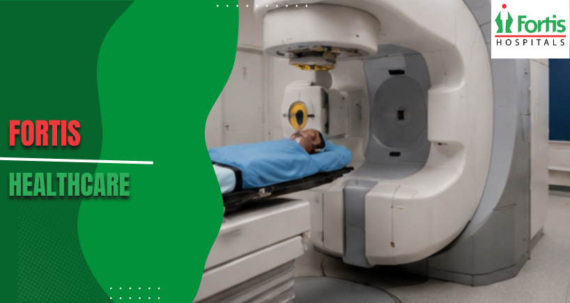
The actual gamma knife surgery procedure takes between 30 minutes to several hours depending on tumour size and location. Patients lie on a treatment bed that slides into the machine, positioning the head frame precisely under the radiation source. The procedure causes no pain because radiation beams produce no sensation when passing through tissue.
Most people remain awake throughout treatment and can communicate with staff through an intercom system. The machine makes clicking sounds as it repositions between beam angles. The Best Hospital in India provides continuous monitoring during procedures to ensure patient comfort and safety throughout the session.
Understanding Treatment Effects on Tumours
Gamma ray surgery damages tumour cells gradually rather than removing tissue immediately like traditional surgery. Radiation disrupts the cellular machinery that allows tumour cells to divide and multiply. These damaged cells die off over weeks to months following treatment.
Imaging scans performed at regular intervals track how tumours respond to radiation exposure. Some tumours shrink noticeably whilst others simply stop growing. The treatment goal focuses on controlling tumour progression rather than achieving instant size reduction.
Managing Common Side Effects
Mild headache and fatigue occur in many patients during the first few days after treatment. These symptoms typically resolve without specific intervention within a week. Some people experience temporary scalp tenderness or minor swelling where the head frame is attached.
Nausea happens occasionally and responds well to standard anti-nausea medications. Hair loss remains minimal because radiation exposure to the scalp stays low. Serious complications occur rarely but may include swelling around the treatment site or new neurological symptoms requiring medical evaluation.
Recovery Timeline and Follow-Up Care
The head frame comes off immediately after treatment completion, allowing patients to return home the same day. Normal activities can usually resume within 24 to 48 hours. Strenuous exercise and heavy lifting should wait for about one week.
Follow-up appointments include brain scans at three-month intervals initially, then extending to six-month or yearly intervals. These imaging studies track treatment effectiveness and detect any tumour changes. The medical team adjusts monitoring schedules based on individual tumour behaviour and response patterns.
Patients should understand that gamma knife treatment represents disease control rather than immediate cure in many cases. Ongoing imaging surveillance remains essential for years, particularly for slow-growing tumours that may change subtly over time. Stable imaging findings often signal treatment success even when tumour size does not visibly decrease, reinforcing the importance of long-term follow-up.
Conditions Treated Beyond Brain Tumours
This technology addresses various brain conditions beyond tumour treatment. Arteriovenous malformations respond well to focused radiation that gradually thickens abnormal blood vessel walls. Trigeminal neuralgia, a severe facial pain condition, often improves when radiation targets the affected nerve.
Acoustic neuromas and pituitary adenomas represent other common treatment targets. Each condition requires specific dose calculations and targeting strategies. The non-invasive nature makes this approach particularly valuable for patients with multiple medical problems who cannot tolerate traditional surgery.
Comparing Effectiveness with Traditional Surgery
Traditional surgery immediately removes visible tumour tissue but requires skull opening and carries risks like bleeding, infection, or neurological damage. Gamma knife surgery in India avoids these surgical complications whilst achieving similar tumour control rates for appropriately selected cases.
Studies show comparable long-term outcomes between both approaches for many tumour types. Patient selection remains crucial because some tumours respond better to one method over another. Tumour size, location, and tissue type all influence which treatment offers optimal results.
Cost Considerations and Insurance Coverage
Treatment costs vary based on tumour complexity and required planning time. Most health insurance plans cover this procedure when medical necessity gets established. Patients should verify coverage details and obtain preauthorisation before scheduling treatment.
Some facilities offer package pricing that includes consultation, imaging, planning, treatment, and initial follow-up visits. Financial counsellors can explain payment options and help arrange insurance approvals. Government schemes may provide coverage for eligible patients meeting specific criteria.
Selecting Appropriate Candidates for Treatment
Ideal candidates have small to medium-sized tumours in locations where traditional surgery poses high risks. Previous surgical attempts that left residual tumour tissue represent another common indication. Patients unable to tolerate general anaesthesia due to medical conditions often benefit from this approach.
Age alone does not determine eligibility because the procedure places minimal physical stress on patients. Doctors evaluate overall health status, tumour characteristics, and treatment goals when recommending this option. Multiple tumours can sometimes receive treatment during a single session.
Technology Advances Improving Precision
Modern systems incorporate real-time imaging that tracks head position throughout treatment delivery. This monitoring compensates for tiny movements that might affect accuracy. Dose calculation software continues improving, allowing even more precise radiation distribution patterns.
Newer machines reduce treatment times whilst maintaining the same accuracy levels. Some centres now offer frameless systems using alternative positioning methods. These advances expand treatment possibilities whilst maintaining the fundamental principle of focused radiation delivery to targeted tissue.
Categories
Clear allRelated Blogs
View all









FAQs

Keep track of your appointments, get updates & more!

