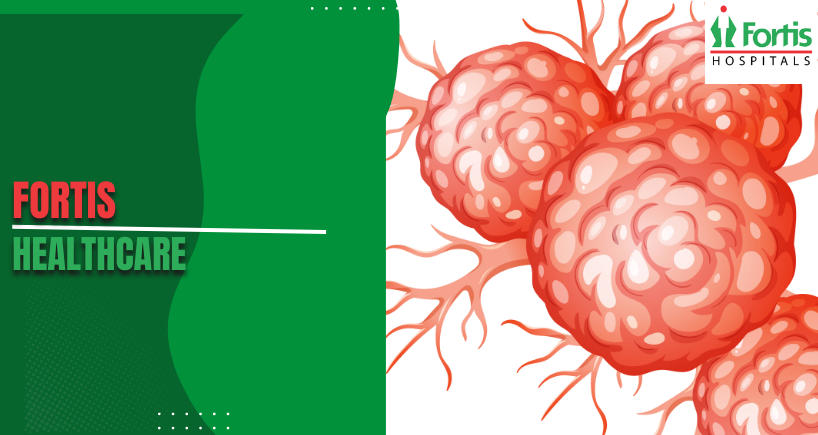


How Neuroblastoma Causes Tumor Growth in Nerve Cells
Cancer arises from complicated biological events which change healthy cells into cancerous ones. Better treatments are being devised by researchers who are working to understand these events and the particular weaknesses that can be targeted. Neuroblastoma originates in early development when specialised nerve cell precursors do not mature as they should.
This cancer of development begins in neural crest cells as they move through the embryo. Normally, these cells become different types of tissue – notably parts of the sympathetic nervous system. However, when the maturing process goes wrong, the immature cells grow out of control instead of turning into working nerves.
Where Neural Crest Cells Come From
Neural crest cells develop alongside the forming spinal cord during the first stages of an embryo’s growth. These remarkable cells travel widely to various places in the body, along specific routes. The cells that become the sympathetic nervous system travel to areas next to the spine. Their eventual locations include the adrenal medulla, sympathetic ganglia and other neuroendocrine tissues.
This movement happens during particular periods of development and is controlled by complicated signalling. Cells must get to the correct places and then receive signals that start differentiation. This complex process involves hundreds of genes working together to control what cells do. Problems at any point can lead to neuroblastoma.
Why Timing of Development is Important
Neuroblastoma in children is a result of development being disrupted during foetal life or in early infancy. The tumours grow from cells that have stopped at an early stage of differentiation. These basic cells keep their ability to multiply instead of reaching full differentiation. The timing of development explains why this cancer mainly affects very young children.
Cells that are going to become part of the sympathetic nervous system have certain transcription factors. These proteins control the programmes of gene expression which cause normal differentiation. Mutations that disturb these regulatory networks allow cells to continue to divide without maturing. The resulting population of cells expands, keeping characteristics from the embryo.
Genetic Mutations as Causes
Most neuroblastoma cases are the result of random, spontaneous genetic mutations. These somatic mutations only affect the tumour cells and are not passed down through families. DNA damage builds up during cell division through mistakes in replication. Unlike many cancers in adults, environmental factors seldom contribute to neuroblastoma.
Some specific genes are repeatedly mutated in tumours from different patients. The ALK gene makes a receptor tyrosine kinase which encourages cell growth and survival. Mutations that activate this protein make it permanently on, whatever the usual regulatory signals. Around 10% of neuroblastomas have ALK mutations that drive multiplication.
The Effect of MYCN Amplification
The MYCN oncogene is vitally important in normal neural development. This transcription factor controls many genes which control cell division and differentiation. Amplification makes hundreds of extra copies of the MYCN gene inside tumour cells. The resulting overproduction of the protein causes very aggressive, uncontrolled growth.
MYCN amplification happens in about 20% of neuroblastoma cases. Tumours with this change behave very aggressively, even with intensive treatment. The extra MYCN protein forces cells to divide continually. It also stops the differentiation programmes which would normally stop multiplication.
Problems with Cell Cycle Regulation
Normal cells carefully control division through checkpoint mechanisms. These protections stop replication if there is DNA damage or if there are not enough nutrients. Neuroblastoma cancer cells get around these checkpoints in many ways. Mutations in the regulators of the cell cycle allow unchecked progress through the stages of division.
The retinoblastoma protein (pRb) normally holds back the start of the cell cycle. Removing this tumour suppressor by inactivating it removes important growth controls. Similarly, p53 mutations prevent apoptosis – programmed cell death – when cells detect problems. These changes allow damaged cells to live and multiply.
Chromosomal Abnormalities Build Up
Neuroblastoma cells often show large-scale changes in chromosomes, beyond individual gene mutations. Loss of chromosome 1p material happens in about 35% of cases. This deletion removes tumour suppressor genes which would normally stop growth. Gaining chromosome 17q material provides extra copies of genes which promote survival.
These chromosomal changes come from errors during cell division. Improperly separated chromosomes make daughter cells with abnormal DNA. Some cells die from too many abnormalities, but others get growth advantages. Natural selection favours cells with mutations that improve fitness in the tumour’s environment.
Signalling Pathway Failures
Many linked signalling networks control what normal cells do. Growth factor receptors on the cell surface receive signals from outside. These messages pass through chains of proteins to eventually reach the nucleus. Transcription factors then turn specific gene programmes on or off.
Mutations affecting parts of the pathways cause inappropriate signalling to be switched on. The PI3K-AKT pathway promotes cell survival and growth when it is stimulated. Constant activation through mutations makes cells resistant to signals to die. Likewise, changes to the RAS-MAPK pathway drive multiplication regardless of growth factors from outside.
Encouraging Angiogenesis
Growing tumours need a blood supply to deliver oxygen and nutrients. Neuroblastoma includes signals which encourage the formation of new blood vessels. Tumour cells release vascular endothelial growth factor (VEGF) which attracts endothelial cells. These cells form new vessels which go into the expanding tumour. Sufficient blood vessel development allows tumours to increase in size beyond what can be seen under a microscope. Without a blood supply, cells at the centre of a tumour die from a lack of oxygen and nourishment; successful neuroblastomas build strong networks of blood vessels to allow continued growth, and anti-angiogenic treatments are designed to break down this vital system of support.
Immune Evasion Strategies
The immune system usually identifies and destroys unusual cells, which includes early-stage cancers. Neuroblastoma cells, however, evolve ways to avoid being noticed by the immune system. They display reduced amounts of the surface molecules that T cells use to recognise them, and this lowered visibility lets the tumours avoid being found.
Certain tumours also release substances which suppress the immune system, inhibiting immune cells in the area; these signals prevent the development of a proper immune response against the tumour. As a result, the area around the tumour becomes immunologically “cold”, with little immune cell activity. A grasp of these ways in which tumours avoid the immune system helps with the creation of immunotherapies. Specialist facilities like Fortis Healthcare, Gurgaon, use full treatment plans including new immunological techniques.
Tumour Heterogeneity Development
Each neuroblastoma contains different groups of cells which are genetically varied. This variety develops from the continual build-up of mutations as the tumour grows. Different groups of cells have different traits, including sensitivity to drugs. Genetic variety allows tumours to change when they are put under the strain of treatment.
This variety makes treatment more difficult as it is hard to destroy every group of cells. Groups of cells which are resistant to treatment survive and eventually cause the illness to return. An understanding of how these groups of cells evolve within tumours helps to create combined treatment plans; by attacking multiple weaknesses at the same time, the development of resistance can be prevented.
Microenvironment Interactions
Tumours exist in complex tissue environments, including stromal cells and immune system components. The behaviour of tumours is influenced by the surrounding cells through several interactions. Fibroblasts release growth factors which help cancer cells to multiply. Immune cells may attack tumours, or – unexpectedly – help them to grow.
The extracellular matrix offers both physical structure and biochemical signals. Cancer cells change this matrix, creating routes for invasion. Blood vessels provide nutrients and also possibly routes for the cancer to spread. Understanding these interactions reveals potential targets for treatment which are not the cancer cells themselves.
Metastatic Mechanisms
Neuroblastoma in children often spreads to distant parts of the body, including the bone marrow and bones. Metastasis – the spread of cancer – requires several coordinated steps, usually known as the invasion-metastasis cascade. First, cells must invade locally through the surrounding tissues. Then, entering the blood or lymphatic vessels allows them to spread to distant locations.
Circulating tumour cells must survive the harsh conditions of the bloodstream. Most cells die, but a few survivors move into distant organs. For growth to be established in these secondary locations, the cells need to adjust to new tissue environments. Only cells which gain the necessary capabilities will successfully establish colonies in distant locations.
Bone Marrow Tropism
Neuroblastoma shows a particular attraction to bone marrow environments. Chemokine receptors on tumour cells guide them toward bone marrow niches. These specialised microenvironments provide growth factors which help cancer cells to survive. Cells in the bone marrow may protect tumour cells from the effects of chemotherapy.
The presence of cancer in the bone marrow is a significant prognostic factor, as it shows widespread disease. Finding cancer in the marrow requires aspiration and biopsy procedures. Monitoring for minimal residual disease uses sensitive techniques to find rare cancer cells. Complete removal of cancer from the marrow is linked to better results.
Differentiation Therapy Potential
Because neuroblastoma develops from halted differentiation, encouraging maturation is a possible treatment. Derivatives of retinoic acid promote the differentiation of neuroblastoma cells into neurons. These differentiated cells stop dividing and may eventually die. Differentiation therapy is part of the standard treatment for patients at high risk.
Not all neuroblastomas react equally to differentiation signals. Genetic features affect sensitivity to these substances. Combining differentiation therapy with other treatments makes it more effective. This approach makes use of the developmental origins of neuroblastoma in a specific way.
Spontaneous Regression Biology
Remarkable spontaneous regression occurs in some infant neuroblastomas. These tumours disappear without treatment, through mechanisms which are not known. The immune system may recognise and destroy cancer cells in these cases. Or, the cells may spontaneously differentiate into harmless tissue.
Stage MS disease in infants, in particular, shows this behaviour. The biological features which underlie regression are not fully understood. Studying these cases gives insights into natural anti-tumour mechanisms. Using these processes therapeutically could help other patients.
Genetic Predisposition Factors
Although most cases happen randomly, some families show a hereditary predisposition. Mutations in the ALK gene increase the risk of neuroblastoma substantially. Mutations in the PHOX2B gene also make this cancer more likely. These inherited cases account for about 1 to 2 percent of all cases.
Hereditary neuroblastoma often appears at younger ages than non-inherited disease. Affected families may have several relatives with cancer. Genetic testing identifies people who carry the mutations and need enhanced monitoring. Understanding predisposition genes reveals critical pathways in tumour development.
Categories
Clear allRelated Blogs
View all


FAQs

Keep track of your appointments, get updates & more!

