


Iridodialysis and Eye Trauma: When the Iris Loses Its Anchor
The human eye absorbs extraordinary physical impacts by utilizing a complex hydraulic system that immediately distributes blunt force evenly across its spherical structure. This defense mechanism has limits. When a sudden, violent compression strikes the globe directly, the rapidly shifting internal fluids create immense shockwaves that violently tear delicate internal tissues apart. The colored portion of the eye represents one of the most mechanically vulnerable structures during these sudden compressions.
Understanding the specific mechanical forces involved clarifies exactly why sudden vision changes require immediate, highly specialized microscopic evaluation. Torn tissue means severe trouble. Within emergency Ophthalmology, recognizing how the internal structures detach from their microscopic moorings dictates the entire surgical approach for vision restoration. A torn iris signifies massive internal disruption that requires far more than simple external observation to protect the patient's long-term sight.
The Mechanics of Blunt Force Tearing the Iris Root
The beautiful colored ring that regulates light entry is extremely thin and anchors itself precisely to the inner muscular wall of the eye. Sudden impacts change the internal geometry. When a high-velocity object, such as a sports ball or snapped elastic band, compresses the cornea backward, the internal hydraulic pressure spikes instantly. This explosive pressure wave forces the iris base to physically rip away from its delicate circular anchor point entirely.
This specific traumatic separation creates a second, artificial opening at the very edge of the colored ring where light can suddenly pour through. The pupil loses its shape completely. Patients look in the mirror and notice that their normally perfectly round pupil has become visibly distorted into an unnatural, flattened shape. Identifying this exact iridodialysis eye injury immediately alerts the examining physician that the eye has sustained a profound mechanical shock.
Experiencing the Immediate Visual Symptoms of Detachment
The sudden creation of a second pupil fundamentally destroys the eye's ability to precisely regulate incoming environmental light levels. The glare becomes completely unbearable instantly. Patients immediately experience agonizing photophobia, where even standard indoor lighting causes sharp, overwhelming physical pain deep within the injured globe. The torn structure flaps loosely inside the fluid-filled chamber, casting confusing, mobile shadows directly across the sensitive retinal tissue located behind it.
The brain receives wildly conflicting visual information that it cannot process because light now enters the eye from two completely different angles simultaneously. This optical confusion generates profound double vision out of a single eye. This bizarre sensory experience leaves the patient intensely disoriented and extremely nauseated within minutes. Managing these overwhelming sensory disruptions requires patching the injured eye entirely just to provide the brain with immediate, necessary relief.
The Hidden Danger of Secondary Bleeding and Pressure
While the torn iris looks highly dramatic externally, the microscopic bleeding triggered by the trauma represents a significantly more dangerous immediate threat to vision. Blood pours directly from the torn root vessels into the clear fluid filling the front chamber of the delicate eye. This thick cellular debris rapidly clogs the microscopic internal drainage system. This physical blockage prevents normal fluid from escaping the globe efficiently.
Internal pressure skyrockets rapidly when the drainage system completely fails, physically crushing the delicate optic nerve located at the very back of the eye. Ophthalmic surgeons repair these delicate torn structures using specialized microscopic suturing techniques that restore vital visual function. Controlling this sudden, massive pressure spike pharmaceutically remains the absolute highest clinical priority before any structural repairs can even be considered safely.
Evaluating the Extent of the Associated Internal Trauma
A force powerful enough to rip the iris from its root almost universally causes significant secondary structural damage deeper within the spherical globe. The trauma rarely stays isolated. Specialists must dilate the pupil fully to search for microscopic retinal tears that could rapidly evolve into massive, blinding detachments. The natural lens sitting directly behind the iris also frequently dislocates during the initial impact, severely compounding the patient's immediate visual distortion.
Clinicians utilize advanced ultrasound imaging to inspect the deepest chambers of the eye when severe internal bleeding completely obscures their direct line of sight. This comprehensive diagnostic sweep ensures no hidden catastrophic damage remains untreated while the surgical team focuses primarily on repairing the visible iris tear trauma. Rushing to fix the cosmetic appearance without confirming the integrity of the retina often results in permanently lost vision.
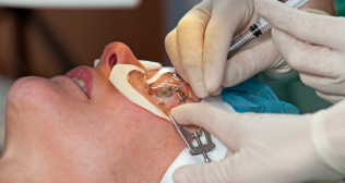
Surgical Techniques for Re-Anchoring the Torn Tissue
Small detachments hidden perfectly beneath the upper eyelid frequently require absolutely no surgical intervention if they cause no visual symptoms whatsoever. However, massive tears demand action. Severe detachments that create unbearable double vision or severe cosmetic deformity demand highly intricate microscopic reconstructive procedures inside the operating room. Surgeons use incredibly fine instruments to gently pull the torn, floating tissue back toward its original anatomical attachment point against the outer wall.
The surgeon carefully sews the delicate colored tissue directly back into the muscular wall using microscopic synthetic threads significantly thinner than a human hair. These complex maneuvers occur within a space barely a few millimeters deep, requiring extraordinary surgical precision to avoid touching the fragile clear lens nearby. Successful suturing eliminates the secondary pupil instantly, completely restoring the eye's natural ability to regulate incoming light efficiently and safely.
The Long-Term Consequences of Severe Anterior Trauma
The traumatized eye remains significantly vulnerable to specific chronic complications for the remainder of the patient's life even after a flawless surgical reconstruction. The microscopic drainage channels damaged during the initial explosive impact frequently develop aggressive scar tissue that slowly chokes fluid outflow over many years. This delayed scarring triggers traumatic glaucoma, a completely painless but devastatingly progressive condition that destroys peripheral vision entirely if left untreated.
Patients who suffer this specific type of violent blunt eye injury require mandatory pressure checks with their specialist at least annually permanently. Wearing highly protective polycarbonate sports goggles becomes absolutely non-negotiable for all future athletic or high-risk industrial activities. Acknowledging that the eye possesses permanently compromised internal architecture ensures patients maintain the strict vigilance required to protect their remaining sight across the ensuing decades.
Categories
Clear allRelated Blogs
View all


FAQs
Can a severely torn iris ever heal itself completely back together without any surgical intervention?
The torn tissue floats suspended in clear fluid, meaning it absolutely never reattaches or heals itself without physical surgical suturing.
Will the injured eye look completely normal again after a successful microscopic reconstructive surgery?
Surgeons restore excellent functional architecture perfectly, but the pupil often remains slightly irregular or subtly flattened upon close cosmetic inspection.
How quickly must the surgical repair be performed after the initial traumatic impact occurs?
Surgeons typically wait several weeks for the acute inflammation and severe internal bleeding to completely resolve before attempting delicate tissue reconstruction.
Are specialized contact lenses effective at treating the severe double vision caused by the tear?
Specialized prosthetic contact lenses painted to match the iris can successfully block the extra light, frequently eliminating the need for complex surgery.
Does the microscopic synthetic thread used to sew the iris remain inside the eye forever?
The incredibly fine synthetic sutures are permanently embedded within the eye wall and never require future extraction or specialized maintenance.

Keep track of your appointments, get updates & more!

