
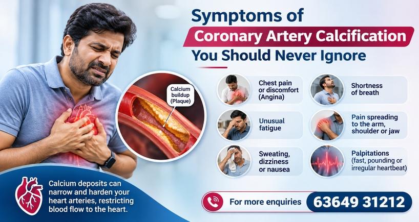


Recognising the Symptoms of Severe Coronary Artery Disease
Introduction
Heart disease is one of the leading health concerns worldwide, and coronary artery disease remains a major cause of cardiovascular complications. While angioplasty is a well-established treatment for blocked heart arteries, certain cases can be particularly challenging, especially when the arteries have severe calcium deposits.
Calcium buildup in the coronary arteries can make them stiff, narrow and difficult to treat using conventional angioplasty techniques. In such situations, image-guided complex coronary angioplasty with calcium modification offers an advanced treatment approach that helps cardiologists assess the blockage more precisely, prepare the artery and optimise stent placement.
With advanced imaging technologies and specialised calcium modification techniques, complex coronary interventions can be performed with greater precision and careful procedural planning.
What Is Image Guided Complex Coronary Angioplasty?
Coronary angioplasty is a minimally invasive procedure used to restore blood flow through narrowed or blocked coronary arteries. During the procedure, a thin catheter with a small balloon is guided towards the blockage. The balloon is inflated to widen the narrowed artery, and a stent is generally placed to help keep the artery open.
In complex cases, particularly those involving heavily calcified arteries, conventional angioplasty may not be sufficient.
Image-guided angioplasty uses advanced intravascular imaging technologies, such as:
- Intravascular Ultrasound (IVUS): Uses ultrasound waves from within the artery to assess the blockage, vessel size and calcium deposits.
- Optical Coherence Tomography (OCT): Uses light-based imaging to provide detailed views of the coronary artery and help assess plaque characteristics and stent positioning.
These technologies help interventional cardiologists understand the anatomy of the artery beyond what can be seen through conventional angiography.
Why Does Calcium Buildup Make Angioplasty Difficult?
Over time, calcium deposits can accumulate within the walls of coronary arteries as part of atherosclerosis, a condition in which plaque builds up inside the blood vessels.
When calcium deposits become extensive, the arteries lose their flexibility. This can create several challenges during angioplasty:
- The balloon may not expand adequately within the narrowed segment.
- The artery may remain rigid despite balloon inflation.
- Stent expansion may be incomplete.
- There may be an increased risk of procedural complications and future stent-related problems.
Therefore, modifying the calcium deposits before stent placement can be an important step in treating selected patients with complex coronary artery disease.
What Is Calcium Modification in Coronary Angioplasty?
Calcium modification refers to specialised techniques used to modify or fracture hard calcium deposits within the coronary artery before placing a stent.
The objective is to make the narrowed artery more flexible, allowing the balloon and stent to expand adequately.
Depending on the patient's condition, the location and severity of calcium deposits, and the imaging findings, interventional cardiologists may use different techniques.
Advanced Calcium Modification Techniques
The choice of technique depends on the patient's coronary anatomy and the treating cardiologist's assessment.
1. Rotational Atherectomy (Rotablation)

Rotational atherectomy uses a specialised catheter with a tiny diamond-coated rotating burr to modify hard, calcified plaque within the artery. It is particularly useful in selected cases involving severely calcified and difficult-to-cross coronary blockages.
2. Intravascular Lithotripsy (IVL)
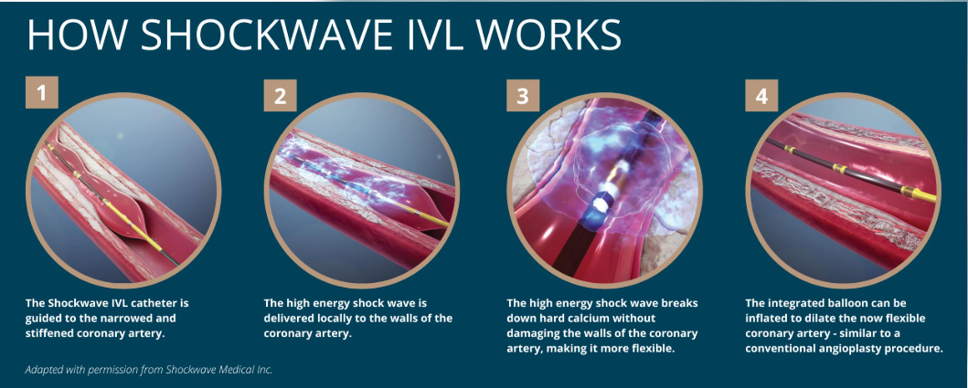
Intravascular lithotripsy uses sonic pressure waves delivered through a specialised balloon to create fractures in calcium deposits within the artery wall. This helps improve vessel compliance and facilitates better stent expansion in appropriately selected lesions.
3. Cutting and Scoring Balloons
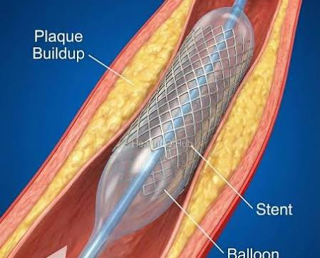
These specialised balloons have small scoring or cutting elements that help modify resistant plaque during balloon inflation. They can be useful in selected lesions where conventional balloon angioplasty does not provide adequate expansion.
The choice between these techniques is based on the extent, thickness and distribution of calcium deposits, along with the patient's overall clinical condition. In certain complex cases, more than one calcium modification technique may be required.
How Does Image Guided Complex Coronary Angioplasty Work?
The procedure involves a carefully planned approach to identify the blockage, modify the calcium deposits and optimise stent placement.
Step 1: Detailed Assessment of the Coronary Arteries
The cardiologist evaluates the coronary arteries using angiography and, where appropriate, advanced imaging techniques such as IVUS or OCT.
Step 2: Identifying Calcium Deposits
The imaging helps determine the extent, thickness and distribution of calcium deposits, allowing the cardiologist to select an appropriate treatment strategy.
Step 3: Calcium Modification
Specialised devices such as rotational atherectomy, intravascular lithotripsy or scoring balloons may be used to modify the calcium deposits.
Step 4: Balloon Angioplasty and Stent Placement
Once the artery has been adequately prepared, a balloon is used to expand the narrowed segment and a stent is placed to help maintain blood flow.
Step 5: Final Imaging Assessment
IVUS or OCT may be used again to assess stent expansion, apposition and the final procedural result.
This imaging-guided approach helps the cardiologist assess the quality of treatment at different stages of the procedure.
PubMed Central (PMC)+1
Who May Require Image Guided Complex Coronary Angioplasty?
This treatment approach may be considered for patients with:
- Severe coronary artery calcification.
- Complex or long-standing coronary artery blockages.
- Narrowed arteries that do not expand adequately with conventional balloon angioplasty.
- Previously stented arteries with underexpanded stents due to calcium.
- Complex coronary artery disease requiring specialised intervention.
- Selected patients with diabetes, kidney disease or other cardiovascular risk factors who have significant coronary calcification.
Not every patient with coronary artery disease requires calcium modification. The treatment approach is determined following a detailed clinical evaluation.
What Are the Potential Benefits?
When appropriately selected and performed by experienced interventional cardiologists, image-guided complex coronary angioplasty with calcium modification can offer several advantages:
- Better procedural planning: Advanced imaging provides a detailed understanding of the coronary artery and the extent of calcium deposits.
- Improved vessel preparation: Calcium modification helps prepare rigid arteries for balloon expansion and stent placement.
- Optimised stent expansion: Helps address one of the important challenges associated with heavily calcified coronary lesions.
- Precision in treatment: IVUS and OCT provide additional information to guide treatment decisions.
- Individualised treatment: Different calcium modification techniques can be selected according to the patient's coronary anatomy.
However, like all cardiac interventions, the procedure carries potential risks, including bleeding, blood vessel injury, coronary artery dissection, heart attack, kidney-related complications and other procedural complications. Outcomes vary depending on individual patient factors.
Recovery After Complex Coronary Angioplasty
Recovery following complex coronary angioplasty depends on the patient's overall health, the complexity of the procedure and whether the intervention was performed as an emergency.
Patients may be monitored in the hospital following the procedure to assess their heart function, blood pressure and recovery.
The treating cardiologist may recommend:
- Taking prescribed medications, particularly blood-thinning or antiplatelet medicines, as directed.
- Following a heart-healthy diet.
- Gradually resuming physical activities based on medical advice.
- Managing diabetes, blood pressure and cholesterol.
- Avoiding smoking and tobacco consumption.
- Attending regular cardiac follow-up appointments.
Patients should never discontinue prescribed antiplatelet medication without consulting their cardiologist, as doing so may increase the risk of stent-related complications.
Why Timely Evaluation of Coronary Artery Disease Matters
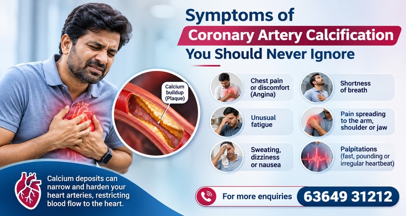
Coronary artery disease can progress gradually, and symptoms should not be ignored. Common warning signs include:
- Chest pain, tightness or discomfort.
- Breathlessness during physical activity.
- Unexplained fatigue.
- Discomfort spreading to the arms, shoulders, jaw or back.
- Sweating, dizziness or nausea associated with chest discomfort.
Some patients, particularly those with diabetes, may experience less typical symptoms.
If you experience sudden chest pain, severe breathlessness or symptoms suggestive of a heart attack, seek emergency medical attention immediately. Do not wait for a routine appointment.
Advanced Cardiac Care at Fortis Hospital, Bannerghatta Road
Managing complex coronary artery disease requires detailed evaluation, appropriate imaging, advanced interventional techniques and individualised treatment planning.
At Fortis Hospital, Bannerghatta Road, patients can consult the Cardiology and Interventional Cardiology team for evaluation and management of coronary artery disease.
If you or your family member has been diagnosed with coronary artery blockages or severe coronary calcification, timely consultation with an interventional cardiologist can help determine the most appropriate treatment options.
Your heart deserves timely attention. Do not ignore the warning signs.
For appointments and enquiries: 63649 31212
Categories
Clear allMeet the doctor

- Cardiac Sciences | Interventional Cardiology
-
 11 Years
-
 1200
Related Blogs
View all







FAQs

Keep track of your appointments, get updates & more!

