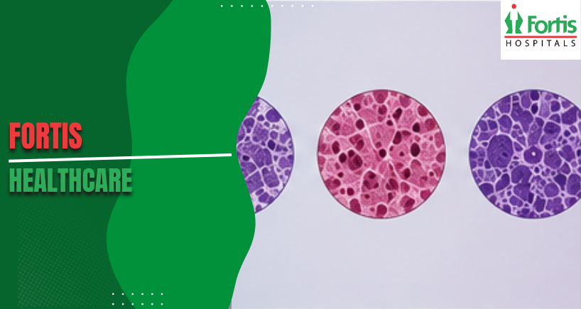


Rhabdomyosarcoma Types: Alveolar vs Embryonal vs Pleomorphic
It isn’t simply paperwork to classify a tumour by its histological subtype; with rhabdomyosarcoma, the subtype determines how aggressively the disease will act, which chemotherapy is best supported by evidence, and what treatment goals are reasonable from the start. Two patients of the same age, and with a cancer beginning in the same place, could have completely different clinical courses solely depending on the subtype their biopsy shows.
This is the reason why rhabdomyosarcoma types represent perhaps the most important classification decision in childhood and adult soft tissue oncology. Treating all three subtypes with the same treatments gives demonstrably worse results than planning treatment according to subtype. What makes these three forms different, at a biological level, is important to all patients, their families, and the doctors involved in managing the disease.
Why Subtype Classification Shapes Every Treatment Decision
The three rhabdomyosarcoma cancer subtypes are not only different in how their cells look under a microscope, but also in their molecular causes, the areas of the body they favour, how they spread, and how they respond to systemic therapy. These aren’t small differences within a single disease; they are clearly separate biological conditions that share a common cellular origin but act very differently clinically once they have developed.
Biopsy should therefore include molecular and cytogenetic analysis, as well as usual histological review. Only looking at appearance creates uncertainty in classification, and this directly leads to uncertainty in treatment. The presence of the PAX-FOXO1 fusion gene, the mitotic index, and how far the cells have differentiated all help to confirm subtype in ways that looking at the tumour’s structure cannot do without molecular testing.
Embryonal Rhabdomyosarcoma and Why It Dominates Paediatric Cases
Embryonal rhabdomyosarcoma is the most common subtype in total, and makes up most of all cases diagnosed worldwide. It mainly affects young children, with the most cases appearing between two and six years old. The tumour most often begins in the head and neck, the eye socket, and the urinary and reproductive systems – places where immature mesenchymal cells become cancerous during early development.
- At chromosome level, embryonal rhabdomyosarcoma usually shows loss of heterozygosity on chromosome 11p15, rather than the specific translocations seen in alveolar histology.
- This molecular profile is linked to comparatively better sensitivity to chemotherapy.
- Standard VAC chemotherapy; vincristine, actinomycin-D, and cyclophosphamide – gives reliable responses in localised disease, and underlies the improvements in survival for rhabdomyosarcoma in children seen over many decades of clinical trial work.
Alveolar Rhabdomyosarcoma and Its More Aggressive Biology
Alveolar rhabdomyosarcoma is less common than embryonal, but much more aggressive biological condition. It happens more frequently in the arms and legs and trunk, occurs across a wider age range including teenagers and young adults. It has a greater possibility to involve local lymph nodes early, and to spread to distant sites when first diagnosed.
The molecular signature of alveolar rhabdomyosarcoma is its most clinically important feature. Most alveolar tumours carry either a t(2;13) or t(1;13) chromosomal translocation, making PAX3-FOXO1 or PAX7-FOXO1 fusion proteins which drive uncontrolled growth and resist the usual signals to cause cell death.
Tumours with these fusions have a worse outlook than those without, whatever the stage; therefore molecular testing, not just appearance, now directs risk assessment in clinical practice.
Pleomorphic Rhabdomyosarcoma as a Distinct Adult Disease
Pleomorphic rhabdomyosarcoma is in a separate clinical category from the two subtypes above. It nearly always occurs in adults – usually patients who are middle-aged or older – and most often starts in the deep soft tissues of the arms, legs and body. Even within the uncommon group; rhabdomyosarcomas in adults, this form is rare; each year, it makes up a small number of all adult soft tissue sarcoma cases diagnosed.
The way the cells are arranged in pleomorphic rhabdomyosarcoma is very unlike the arrangement in embryonal or alveolar rhabdomyosarcoma. The tumour has extremely varied, unusual, large cells containing multiple nuclei, and doesn’t show the organised patterns of growth seen in the other types.
This lack of cellular order is linked to aggressive behaviour of the disease, a greater chance of spreading to distant parts of the body when first found, and a poorer reaction to the chemotherapy treatments which do well in the kinds of rhabdomyosarcoma which are most often seen in children.
Diagnostic Pathway That Accurately Separates the Three Subtypes
To correctly identify the subtype, a planned series of diagnoses is needed which combines scans, tissue samples, and molecular tests. Hospitals deal with this through specialist sarcoma pathology groups who have access to immunohistochemistry tests and molecular testing systems to give a certain identification of the subtype. This set-up is particularly important when the shape of the tumour cells is not clear, or when the status of fusion genes needs to be confirmed before treatment can be planned.
Before a biopsy, an MRI scan of the place where the tumour first developed is done in every instance, to establish how large the tumour is and to show the safest way to take a sample. A core needle biopsy – not removal of the whole tumour – is the usual method, protecting the chances of surgery while getting enough tissue for both shape and molecular tests. To remove the tumour before properly finding the subtype harms later staging and the plan for treatment in ways that are hard to put right later.
How Rhabdomyosarcoma Symptoms Differ Across Subtypes
Rhabdomyosarcoma symptoms change a lot, depending on the subtype and where the first tumour has grown. Embryonal tumours in the head and neck cause bulging of the eye, blockage of the nose, or a visible swelling in the neck of young children. Alveolar tumours in the arms and legs appear as firm, increasing swellings of the limbs, with associated stiffness or less movement in joints as the growth becomes larger within muscle areas.
Pleomorphic tumours in adults often produce large growths in the retroperitoneal area or the trunk, with unclear pain in the stomach, pressure on the side, or urine blockage, before the main growth can be felt directly. This difference in symptoms shows why rhabdomyosarcoma symptoms can’t be judged without knowing where in the body they are, and why a soft tissue growth which stays for over three weeks without a known harmless reason needs a scan, not continued medical watching.
Treatment Intensity That Reflects Each Subtype's Biology
- Rhabdomyosarcoma in adults with alveolar or pleomorphic structure needs much more intensive treatment planning than embryonal cases in children with disease limited to one area. Alveolar cases get stronger chemotherapy, wider checking of lymph nodes, and radiotherapy to nearby areas, whatever the state of surgical edges. Pleomorphic cases in adults are frequently treated using adult soft tissue sarcoma methods instead of children’s systems, because of the different biology and the different ability of organs to stand treatment.
- Rhabdomyosarcoma in children with embryonal structure and completely removed disease limited to one good place is at the other end of the scale of treatment intensity. Here, the main clinical aim is to achieve a cure while reducing long-term harm from treatment in a growing child. The three subtypes of rhabdomyosarcoma at last need three very different clinical talks about what treatment means, what result is expected, and what long-term checking will need from both the patient and the team treating them.
Categories
Clear allRelated Blogs
View all


FAQs

Keep track of your appointments, get updates & more!

