


Understanding Symblepharon: Causes, Types and Treatment Options
The conjunctiva is a thin, clear membrane that covers the white part of your eye (the sclera) and lines the inside of your eyelids. Symblepharon (plural: symblephara) occurs when these two layers of the conjunctiva become stuck together.
Symblepharon happens when strong inflammation causes the tissues in your eye to swell and stick together. As this continues, scars can form, causing the layers to fuse together permanently. You might have one or more adhesions. If the inflammation in your eye persists, it can lead to repeated episodes of symblepharon.
Symptoms
A symblepharon is often visible in the eye as a band of tissue connecting the inner eyelid to the eyeball. If you cannot see it directly, you may notice that your eyelid doesn’t open fully or that your eyeball has limited movement. Other possible symptoms of symblepharon include:
- Ptosis: Symblepharon can cause your eyelid to droop by pulling it downward.
- Entropion: It may cause your eyelid to turn inward toward your eye.
- Lagophthalmos: Symblepharon can prevent your eyelid from fully closing.
- Diplopia: Restricted eye movement may lead to double vision.
These can also result in eye irritation and other symptoms, such as:
- Redness
- Sensitivity to light
- Excessive tearing
- Dryness in the eye
Causes
Severe or chronic inflammation of the conjunctiva (conjunctivitis) can result in the development of symblepharon. Symblepharon can arise from external factors like chemical or thermal burns, as well as pterygium, or from internal conditions such as Stevens-Johnson syndrome (SJS), ocular cicatricial pemphigoid (OCP)/mucous membrane pemphigoid (MMP) and dry eye disease. Symblepharon can also develop as a severe side effect of drug reactions or following glaucoma filtering surgery (GFS) due to the denudation of the conjunctival epithelial surface.
Types
There are three types of symblepharon, namely:
- Anterior symblepharon: It occurs when the edge of the eyelid, or a portion of it, becomes adherent to the surface of the eyeball.
- Posterior symblepharon: It occurs when the conjunctival fornix or cul-de-sac is affected.
- Total symblepharon: It occurs when the entire eyelid surface adheres to the globe, resulting in the obliteration of the fornix.
Diagnosis
Your eye care provider can diagnose symblepharon with a detailed eye exam. To help make you more comfortable, they may use eye drops. During the exam, they will carefully check which parts of the eye are affected and where the tissues are sticking together. They will also look for any complications that might be related to the specific type of symblepharon you have.
If they are unsure of what’s causing your symblepharon, your provider may suggest further tests, like an eye swab or blood test, to help pinpoint the cause. Some conditions may improve with treatment, while others might be long-term and require ongoing care.
Treatment
Symblepharon treatment includes the following:
Medications: Preventing symblepharon is usually easier than treating it. If your provider knows you are at risk or have a condition that might cause it, they can take steps to help prevent it from happening. Treatment for the underlying condition may involve:
- Corticosteroids to reduce inflammation
- Antibiotics or antivirals to treat infections
- Immunomodulators for autoimmune diseases
You may be prescribed medications to take orally or as eye drops. After the inflammation in your eye has subsided, your provider may apply an amniotic membrane dressing to help prevent or reduce scarring.
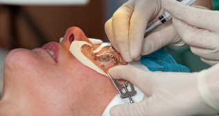
- Surgery: If your provider is unable to prevent symblepharon or is treating it after it has developed, surgery may be needed to remove the adhesions. The type of surgery required will depend on the severity of the condition and include:
- Symblepharon release: This procedure involves separating the adhesions between the layers of your conjunctiva. In some cases, your surgeon may also need to remove scar tissue from the conjunctiva.
- Tissue graft: Surgeons may use tissue from another part of your eye or from a different mucous membrane, such as the inside of your lip, to replace the tissue that is removed.
- Oculoplastic reconstruction: If symblepharon has impacted your conjunctival fornix, your surgeon may need to reconstruct it. If the cornea is affected, keratoplasty (corneal transplant) may be necessary.
Complications
If left untreated, symblepharon can result in various complications, which may vary based on the severity and extent of the adhesions. Possible complications include:
- Visual impairment: In cases where symblepharon leads to substantial adhesions, it can result in a decrease in visual acuity, potentially causing blurred or distorted vision.
- Chronic discomfort: People with symblepharon may experience ongoing discomfort, such as dryness, irritation or the sensation of a foreign object in the eye.
- Corneal complications: In severe cases of symblepharon, the cornea may be affected, potentially resulting in corneal abrasions, ulcers or other corneal issues.
- Conjunctival scarring: The scarring caused by symblepharon can alter the structure of the conjunctiva, affecting tear production and overall eye health.
- Functional impairments: Symblepharon can disrupt the normal function of the eyelids and tear film, leading to conditions like dry eye syndrome.
- Psychosocial impact: The visible effects of symblepharon and its potential impact on appearance can have psychosocial consequences, influencing a person’s self-esteem and body image.
Conclusion
The outcome for symblepharon management can differ significantly based on factors such as the underlying cause, the severity of the adhesions and the effectiveness of the treatment. Generally, early detection and prompt treatment lead to better outcomes.
Categories
Clear allRelated Blogs
View all


Keep track of your appointments, get updates & more!

