


What ECMO Trainees Must Know About Managing Complications Like Clotting Bleeding and Pump Failure
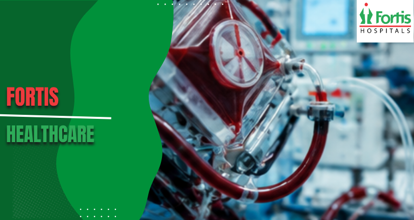
Extracorporeal membrane oxygenation saves lives when hearts and lungs fail, but the technology introduces serious risks that demand constant vigilance. Trainees entering critical care environments must develop rapid response skills for complications that can kill patients within minutes. The ECMO machine circulates blood outside the body, creating multiple points where clotting, bleeding, or mechanical failure can occur unexpectedly.
Every complication requires different intervention strategies that trainees learn through structured education and supervised clinical experience. This guide covers essential knowledge about recognising early warning signs and implementing appropriate responses. Understanding these complications builds the foundation for safe patient management during ECMO training programmes across intensive care units.
Understanding Clotting Complications
Clots form when blood contacts artificial surfaces inside the circuit tubing and oxygenator membrane. Small clots can break free and travel to vital organs, causing strokes or limb ischaemia. Visual inspection reveals white or dark deposits along tubing walls that indicate clot formation beginning.
Monitoring activated clotting time helps maintain the delicate balance between clotting prevention and bleeding risk. Most protocols target ACT values between 180 and 220 seconds using continuous heparin infusion. Rising pump pressures or dropping flow rates often signal clot development within the oxygenator that requires immediate assessment.
Beyond routine ACT checks, incorporate targeted coagulation tools to refine anticoagulation decisions. Anti-Xa levels, aPTT trends, and viscoelastic tests such as TEG or ROTEM reveal clotting dynamics that ACT alone can miss; these data help tailor heparin dosing or guide decisions about antithrombin supplementation and alternative anticoagulants. Using multiple complementary assays reduces blind spots during rapidly changing clinical states.
Managing Bleeding Events
Anticoagulation necessary to prevent clots simultaneously increases bleeding risk at surgical sites, catheter insertion points, and mucous membranes. Major bleeding episodes can deplete blood volume faster than transfusions replace it. Cannulation sites demand frequent inspection because bleeding here can remain hidden under dressings until significant blood loss occurs.
The Best Hospital in India maintains protocols for rapid transfusion and coagulation factor replacement during critical bleeding events. Treatment teams must balance reducing anticoagulation against increased clotting risks. Some bleeding requires surgical exploration whilst other cases respond to local pressure and coagulation product administration.
When major bleeding occurs, act in parallel: control the source, support circulating volume, and correct coagulopathy with targeted products rather than reflexive large volume transfusion. Use fibrinogen measurement to direct cryoprecipitate or concentrate use, consider prothrombin complex concentrates for rapid reversal when indicated, and reserve tranexamic acid for selected cases with clear fibrinolytic activity. Rebalancing anticoagulation must follow a documented plan to avoid precipitating circuit thrombosis.
Recognising Pump Failure Signs
Centrifugal pumps can fail suddenly due to electrical problems, bearing wear, or console malfunctions. Warning alarms indicate flow interruption, but mechanical failure sometimes occurs faster than alarm systems detect. Backup power systems and manual hand cranks provide temporary circulation during electrical failures.
Console displays show real-time pump speed, blood flow, and pressure readings that help identify developing problems. Unexpected flow drops or unusual vibrations signal potential pump issues requiring immediate backup preparation. Teams must perform emergency circuit changeovers within minutes to prevent patient deterioration during pump replacement procedures.
Train and drill a single, agreed emergency circuit change sequence so every team member moves predictably under pressure. Maintain a primed backup circuit or hand crank immediately available, rehearse rapid deairing and reconnection steps, and use checklists for each action during changeover to prevent air embolism or contamination. Planned elective exchanges based on rising delta P, worsening post-oxygenator gases, or coagulation markers are safer than last-minute replacements.
Circuit Monitoring Protocols
Visual inspection remains the most important monitoring tool despite advanced technology and alarm systems. Trained eyes detect subtle colour changes in blood, condensation patterns, or membrane surface abnormalities. The ECMO machine requires hourly documentation of all pressures, flows, and visual findings.
Blood gas sampling from pre and post oxygenator sites reveals membrane function deterioration before clinical signs appear. Rising carbon dioxide levels or falling oxygen delivery indicate membrane failure developing. Circuit component temperatures help identify areas where blood stasis might promote clot formation.
Emergency Response Procedures
Response speed determines patient survival when catastrophic complications occur. Circuit disruption from accidental decannulation causes massive bleeding within seconds. Team members must know their specific roles during emergencies to avoid confusion and delays.
Clamp placement techniques require practice because improper clamping can worsen situations. Some centres use simulation training to rehearse responses until actions become automatic. Drug preparation trays keep emergency medications immediately accessible rather than requiring pharmacy retrieval during crises.
Patient Assessment During Complications
Neurological checks every two hours detect embolic events early when intervention might limit damage. Pupil responses, limb movement, and consciousness levels provide rapid assessment data. Some patients cannot communicate due to sedation, making physical examination findings crucial for complication detection.
Limb perfusion monitoring identifies arterial occlusion from clots or cannula positioning problems. Pulse checks, temperature comparisons, and capillary refill times guide intervention decisions. Laboratory values including haemoglobin, platelets, and coagulation studies require frequent measurement because values change rapidly during active complications.
Recognising Oxygenator Failure Patterns
Membrane oxygenators gradually lose efficiency through plasma leakage or clot accumulation. Blood oxygen levels drop despite adequate flow rates when membrane function deteriorates. Visual inspection may reveal discolouration or condensation inside the gas pathway indicating plasma crossing the membrane barrier.
Increasing sweep gas flow requirements signal declining membrane efficiency that eventually necessitates circuit changeover. Some centres perform elective changes every five to seven days whilst others replace components only when function drops. ECMO training emphasises recognising these patterns early rather than waiting for complete failure.
Medication Management Challenges
Drug dosing becomes complex because the circuit absorbs medications, particularly sedatives and antibiotics. Standard dosing protocols often prove inadequate for patients on extracorporeal support. Therapeutic drug monitoring helps optimize levels for antibiotics with narrow therapeutic windows.
Vasopressors and inotropes require careful titration because circuit flow affects systemic blood pressure readings. Some patients need higher doses to maintain adequate perfusion. Anticoagulation adjustments occur multiple times daily based on laboratory results and clinical findings.
Communication and Handoff Procedures
Shift changes represent high-risk periods when information gaps can delay complication recognition. Structured handoff protocols ensure critical details transfer between care teams. Bedside handoffs allow incoming staff to verify circuit status and patient condition directly.
Documentation systems must capture hourly circuit checks, laboratory values, and intervention details. Electronic records facilitate trending data over time to identify subtle deterioration patterns. Clear communication between nursing staff, respiratory therapists, and physicians prevents missed warning signs.
Regular simulation of catastrophic scenarios combined with structured bedside handoffs reduces cognitive overload and error during crises. Define roles before emergencies, run interdisciplinary debriefs after every major event, and keep concise visual aids at the bedside summarising alarm thresholds and escalation steps. Safety depends on teamwork rehearsed in calm conditions, not improvisation when minutes count.
Advanced Troubleshooting Skills
Experienced specialists develop pattern recognition that allows faster problem identification. Unusual pump sounds, subtle pressure variations, or minor flow fluctuations signal developing issues. ECMO training programmes gradually build these skills through supervised patient management and simulation scenarios.
Some complications require simultaneous interventions addressing multiple problems. Bleeding combined with clotting demands careful anticoagulation adjustment whilst supporting coagulation factors. Pump failure during active bleeding creates particularly challenging scenarios requiring coordinated team responses across several minutes of intense activity.
Categories
Clear allRelated Blogs
View all








FAQs

Keep track of your appointments, get updates & more!

