


ECG vs ECHO Test: Navigating the Differences!
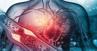
Cardiovascular disease (CVD) is a common term used for conditions affecting the heart or blood vessels (arteries, veins, or capillaries).
The prevalence of cardiovascular diseases (CVD) in India is remarkably higher than what is being experienced at global level. Ischemic heart disease and stroke are the leading causes and are accountable for more than 80% of CVD deaths. Furthermore, 50% heart attacks are experienced by Indian men below the age of 50 years. Warning signs and symptoms of heart attack include chest pain, shortness of breath, and more. Therefore, in dealing with CVDs, an accurate diagnosis is the first critical step to crafting an effective treatment plan.
When it comes to cardiac diagnostics, we often come across terms like electrocardiogram (ECG) or echocardiogram (ECHO). Ah! But are both the terms synonymous or is there a difference? Let us dive deep to understand and obtain better insights about the differences between two terms:
Parameter | ECG test | ECHO test |
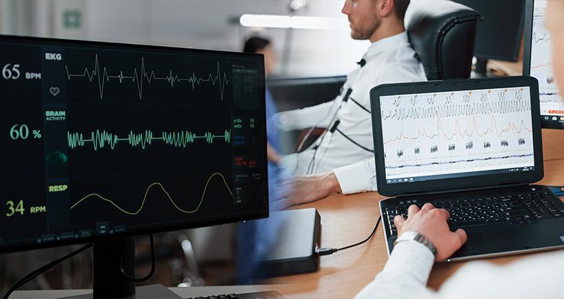
Definition | An electrocardiogram (ECG) is a simplest and useful test which records how often the heart beats, heart rhythm and electrical activity of person’s heart. | An echocardiogram, or “echo” is a scan utilized to glance at the heart and nearby blood vessels. |
What happens during the test? | Ten miniature sticky patches referred to as electrodes are put on person’s arms, legs, and chest. These are connected by lead wires to an ECG machine which takes up the electrical signals which make a person’s heartbeat. This electrical activity is recorded and is printed manually. | During test, healthcare professional utilizes ultrasound (high-frequency sound waves) from a hand-held wand placed on chest to capture pictures of heart’s valves and chambers. This aids the provider evaluate the pumping action of heart. Providers often combine echo with Doppler ultrasound and colour Doppler techniques to assess blood flow across heart’s valves.
|
Time Spent | This procedure normally takes between 5-10 minutes. | It varies from individual to individual and can take from 15 minutes up to an hour. |
Types | Healthcare providers utilize several types of electrocardiograms (ECG) to look for an array of cardiac (heart) conditions. Some of the extensively used types are: Resting ECG: It is a non-invasive test which can detect abnormalities such as arrhythmias, evidence of coronary heart disease, etc. The procedure involves lying still on back with a bare chest. It is significant that person lies calmly and comfortably during the test because tense muscles, moving, coughing, or shaking can impact the results. This type of ECG utilizes about one minute, or five minutes at most for measurement. Exercise ECG: Here the electrical activity of heart is estimated while a person is physically active. This usually involves walking on a treadmill or riding an exercise bike. | An echocardiogram may utilize certain special types of echocardiography, as listed below: M-mode echocardiography: It is the simplest type of echocardiography generates an image which is similar to a tracing rather than an actual picture of heart structures.M-mode echo is helpful for examining heart structures, such as the heart's pumping chambers, the size of the heart itself, and the thickness of the heart walls. Doppler echocardiography: This technique is utilized to estimate and analyse the flow of blood through the heart's chambers and valves. The quantity of blood pumped out with each beat is a sign of the heart's functioning.
|
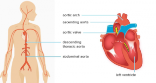
Purpose or application | It can provide us crucial information, for instance about narrowing of the coronary arteries, a heart attack, or an irregular heartbeat such as atrial fibrillation. | An echocardiogram can aid diagnose and monitor certain cardiac conditions. It examines the structure of the heart and surrounding blood vessels, validating how blood flows through them, and assessing the pumping chambers of the heart. An echocardiogram can aid detect damage from a heart attack, heart failure, congenital or birth-related heart disease, heart valve abnormalities, cardiomyopathy, and endocarditis. |
Enormous cutting-edge research into myocardial infarction treatments could save lives and improve the health span of individuals further enhancing quality of life. If a person has particularly high chances or risk of developing CVD due to blood cholesterol or high blood pressure, they should immediately consult a cardiologist or suitable healthcare provider.
Categories
Clear allRelated Blogs
View all


Quick Enquiry Form

Keep track of your appointments, get updates & more!