


Clinical Pearl of the Month: Robotic Precision in Managing a Large Cervical Fibroid
At the Centre for Advanced Gynaecology, Urogynaecology, Gynaec Oncology & Robotic Surgery at Fortis Hospital Bannerghatta Road, complex gynaecological conditions are managed with advanced technology and a patient-centred approach.
In this month’s clinical highlight, Dr. Rubina Shanawaz Z and her team successfully treated a 33-year-old police officer suffering from severe urinary symptoms caused by a large cervical fibroid compressing the bladder. The case demonstrates how robotic-assisted surgery can safely address complex fibroids while preserving the uterus and enabling rapid recovery.
When Fibroids Cause Pressure Symptoms
Fibroids are common benign growths of the uterus, but cervical fibroids account for less than 5% of all fibroids. Due to their location near the bladder, ureters, and rectum, they can present differently from typical uterine fibroids.
Unlike the usual symptoms of heavy menstrual bleeding or pelvic pain, cervical fibroids may produce pressure-related urinary symptoms, often resembling primary bladder disorders.
This case highlights how advanced robotic surgery can provide precise treatment for such challenging cases while maintaining fertility and normal anatomy.
Patient Presentation
The patient, a 33-year-old police officer (P2L2) with a history of two previous caesarean deliveries, presented with a six-month history of urinary symptoms that were significantly affecting her daily life and work responsibilities.
Her symptoms included:
- Increased urinary frequency
- Sensation of incomplete bladder emptying
- Nocturia (waking 3–4 times at night to urinate)
- Intermittent suprapubic discomfort
Despite these symptoms, she reported normal menstrual cycles without heavy bleeding or severe pain, which is unusual for fibroid-related conditions.
Given the physically demanding nature of her profession, the symptoms were interfering with her active duty performance.
Clinical Examination and Diagnosis
During the clinical evaluation, doctors identified a firm pelvic mass palpable above the pubic bone.
Pelvic examination revealed:
- Posterior displacement of the cervix
- A smooth, firm mass arising from the cervix
- Reduced uterine mobility
- Fullness in the anterior vaginal fornix
Further imaging confirmed the diagnosis.
Imaging Findings
Ultrasound Scan
- 8.8 × 9 cm well-defined hypoechoic mass
- Arising from the anterior cervical wall
- Causing compression of the bladder
MRI Pelvis (Fibroid Mapping)
- Large 9 × 8.7 × 8 cm solitary cervical fibroid
- Significant displacement and compression of the bladder
- Bilateral ureters stretched laterally
- No signs of malignancy
Since the patient was young and wished to preserve her uterus, the medical team recommended robotic myomectomy, a minimally invasive surgical approach that removes fibroids while preserving the uterus.
Surgical Plan
The surgical strategy included a stepwise minimally invasive approach:
- Cystoscopy to visualize the bladder and ureteric openings
- Bilateral DJ stenting to protect the ureters during surgery
- Robotic myomectomy to safely remove the fibroid
This approach ensured maximum surgical precision and protection of nearby organs.
Intraoperative Findings
During surgery, doctors observed:
- A large anterior cervical fibroid (FIGO Type 8)
- Distortion of the lower uterine segment
- Bladder displaced forward and adherent to the uterus
- Bilateral ureters displaced laterally
The close proximity of these vital structures made the case technically complex, requiring meticulous surgical planning.
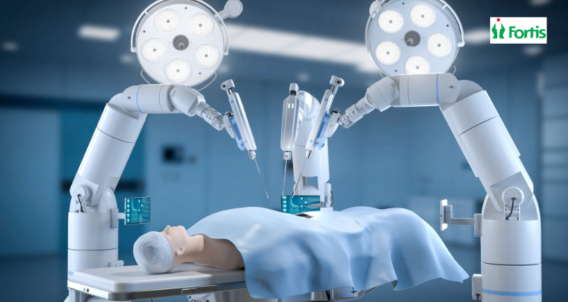
Robotic Surgical Procedure
Using the robotic surgical platform, the surgical team performed the following key steps:
- Visualization and lateralization of the ureters
- Careful dissection of the bladder from the fibroid
- Vasopressin injection to reduce bleeding
- Precise enucleation of the 9 cm cervical fibroid
- Inspection of the endocervical canal
- Layered reconstruction of the uterine muscle
- Hemostasis with minimal blood loss
- Removal of the fibroid using in-bag power morcellation
The robotic system provided enhanced magnified visualization, improved instrument dexterity, and superior suturing capabilities, allowing safe surgery even in this anatomically challenging location.
Smooth Recovery and Excellent Outcome
The patient recovered smoothly under an Enhanced Recovery After Surgery (ERAS) protocol.
Her postoperative course included:
- Discharge on postoperative day 1
- Complete resolution of urinary symptoms
- Improved bladder emptying
- Return to active police duty within two weeks
- Normal menstrual cycles maintained
Histopathological analysis confirmed the diagnosis of benign leiomyoma (fibroid).
Why Robotic Myomectomy Matters
Cervical fibroids can be difficult to manage surgically due to their proximity to the bladder, ureters, and pelvic nerves.
Robotic surgery offers several advantages in such cases:
- High-definition magnified visualization
- Precise dissection around delicate structures
- Minimal blood loss
- Superior uterine reconstruction
- Faster recovery and shorter hospital stay
For young women who wish to preserve fertility and uterine function, robotic myomectomy provides a safe and effective treatment option.
Expertise in Advanced Robotic Gynaecology
This complex surgery was performed by the expert team at the Centre for Advanced Gynaecology, Urogynaecology, Gynaec Oncology & Robotic Surgery at Fortis Hospital Bannerghatta Road.
The team includes:
- Dr. Rubina Shanawaz Z
- Dr. Punyashree R M
- Dr. Prakruthi S
Their expertise in robotic and minimally invasive surgery enables the management of complex gynaecological conditions with precision, safety, and faster recovery.
Dr. Rubina Shanawaz Zameer | Fortis Hospital CG Road Bangalore
Categories
Clear allMeet the doctor

- Obstetrics and Gynaecology | Robotic Surgery | Gynaecologic Oncology | Obstetrics and Gynaecology
-
 16 Years
-
 1150
1150
 Available at 1 different locations
Related Blogs
View all

Bacterial Vaginosis: Causes, Symptoms And Treatment


Awareness, Breast Self-Examination And Regular Screening Examination Can Change The Life o...

Hormonal Imbalance: Symptoms, Causes And Treatment

Evolution of Gynae & Cancer Surgeries With Robotics


Know The Top 11 Benefits of Antenatal Exercises


FAQs

Keep track of your appointments, get updates & more!

