


Olfactory Neuroblastoma: Symptoms, Diagnosis & Treatment Plan
A lot of people do not worry about slight nasal problems, thinking ongoing congestion is due to hay fever or frequent colds which get better by themselves. But, symptoms which get steadily worse for a number of months should have a full medical check by the correct experts. Olfactory neuroblastoma is a very unusual cancer developing from specialist nerve tissue in the upper part of the nose. This rare tumour affects about 400 people yearly, across the world, so most patients – and many doctors – don’t know about it.
This illness is quite different from the typical neuroblastoma in children, as it mainly affects adults aged 40 to 70. The tumour grows slowly, often giving subtle signs which at first are easily thought to be harmless conditions, or wrongly diagnosed. Knowing the usual early warnings, how it is diagnosed, and all the ways it can be treated, allows patients to quickly get the right specialist help.
What Makes Olfactory Neuroblastoma Different From Childhood Neuroblastoma
Neuroblastoma is the name for several separate cancers that all begin in nerve tissue, but behave in very different ways biologically. Childhood neuroblastoma develops from cells which will become part of the sympathetic nervous system, usually in the adrenal glands or along the sympathetic nerve chain. These childhood tumours grow quickly, often showing widespread disease, but surprisingly respond well to treatment.
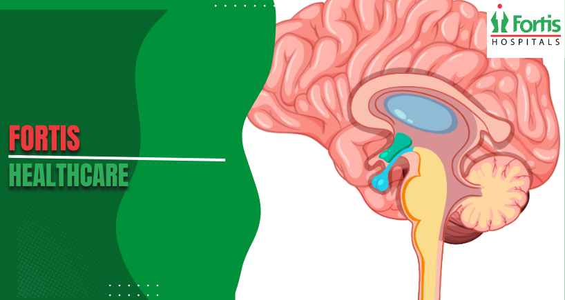
Olfactory neuroblastoma happens in the olfactory epithelium specifically, which lines the upper part of the nasal cavity near the base of the skull. This specialist tissue holds nerve cells which detect smells and send smell signals to the brain. The tumour develops more slowly than the childhood types, sometimes taking years to give symptoms which make a person seek medical advice.
Early Signs – Often Thought to be Sinus Issues
The first signs usually start with nasal obstruction on one side, affecting only one nostril when it first appears. This increasing blockage slowly gets worse over months as the tumour gets larger in the small nasal space. Many people think their symptoms are chronic sinusitis, and try shop-bought decongestants which do not give lasting help. The fact that it’s always on one side should show it is cancer, and not the normal congestion in both nostrils that happens with sinus problems.
Patients often see a number of ordinary doctors before being correctly sent to a specialist for a full examination. The slow development and the fact that the symptoms are the same as harmless conditions cause delays in diagnosis, averaging six to twelve months. If symptoms go on despite normal treatments for sinus issues, a referral to an ear, nose and throat specialist is needed for a full nasal endoscopy examination.
Understanding Smell Loss as a Diagnostic Signal
Olfactory dysfunction happens when tumours harm or fully destroy the nerve endings which detect smell. Most patients notice a gradual loss of smell in the blocked nostril at first, and it may then become in both nostrils. This symptom is not like the temporary loss of smell during a virus infection, as it goes on for ever and does not get better. Many people get used to this without realising how big the loss is, until they are specifically asked about it during medical assessment.
Complete anosmia – total loss of smell – shows that the olfactory nerves have been badly harmed by the tumour directly.
Taste depends a lot on smell, so patients often say their taste is changed along with the smell problems.
These sense changes rarely get fully better, even after the tumour has been successfully treated, and are permanent functional losses.
When Nosebleeds Show Serious Problems
Epistaxis develops as growing tumours break down blood vessels in the very well-supplied nasal mucosa which lines the nose. These bleeding episodes can be from slight spotting to major haemorrhage needing urgent medical help and nasal packing. The bleeds happen by themselves, without obvious causes like nose picking or injury to the face. Repeated episodes despite normal treatment need a full nasal examination by experienced ear, nose and throat specialists.
New blood in nasal discharge or when blowing the nose shows active bleeding from the easily broken surfaces of the tumour. Serious bleeds sometimes need visits to the emergency department for anterior or posterior nasal packing. Anyone over 40 with repeated, unexplained epistaxis should have a full nasal endoscopy, looking at the whole cavity.
Diagnostic Pictures Showing How Much Tumour There Is
Computed tomography scanning gives detailed anatomical information showing the patterns of bone destruction which clearly show how the tumour is invading. High-resolution CT shows erosion of the skull base, involvement of the sinuses, and extension into the eye socket with very good anatomical clarity. Coronal and axial views give a three-dimensional understanding of the tumour’s relationships with nearby critical structures. Intravenous contrast enhancement helps show what is actual tumour tissue, and what is inflammation and normal anatomy.
Magnetic resonance imaging gives better soft tissue detail, which is essential for complete surgical and radiation treatment planning. T1-weighted pictures show anatomical relationships whilst T2-weighted images show how much tumour there is and patterns of surrounding oedema. Gadolinium contrast helps show the edges of the tumour more clearly than pictures without contrast can. MRI is best at showing the tumour going into the skull and involving the dura – which CT imaging might underestimate a lot. Confirmation by Tissue Biopsy is Essential
Accurate diagnosis depends on a microscopic study of tumour tissue, and this is gained through careful biopsy via endoscopy. Depending on where the tumour is, and the patient themselves, ENT surgeons do biopsies with either local or general anaesthetic. Getting enough tissue is important, so pathologists can do a full examination, including specialist immunohistochemistry. The biopsy gives a definite diagnosis, and also important information about how the cancer will behave, by carefully assessing the tumour’s grade.
Pathologists look at the structure of the cells, finding features such as pseudorosette formations, and the neuronal differentiation markers which are specific to neuroblastoma. Immunohistochemistry proves a neural origin, by showing particular protein expression patterns – and this is how it’s distinguished from other tumours. How the tumour is graded affects what treatment is recommended, and helps to forecast how the patient is likely to do, and their prognosis.
Surgical Removal as the Main Treatment
If it’s possible to do so without causing too much harm, removing the entire tumour is the most important part of treatment which offers a cure. In selected cases, endoscopic endonasal surgery lets surgeons remove the tumour through the nostrils, without any cuts on the face. This less invasive technique cuts down recovery time a lot, and doesn’t leave any cosmetic scarring, when compared with traditional open surgery. Using high-definition cameras and endoscopes at specific angles, surgeons get very good views of hard-to-reach areas.
Larger tumours need open craniofacial resection, which combines the knowledge of neurosurgeons and ENT surgeons to remove the tumour safely and completely. This combined approach provides the access needed for tumours which have grown into the skull, or which involve the base of the skull a lot. During tumour removal, the surgical team reconstructs any resulting defects using local or microvascular free tissue flaps. Centres like Fortis Healthcare, Gurgaon, have dedicated programmes for dealing with complex cancers of the head and neck, and which need a team of specialists.
Radiotherapy After Surgery
Radiotherapy after surgery is to treat any microscopic disease which may remain, even after a very good attempt at surgical removal.
Doses are usually between 54 and 66 Gray, given over six or seven weeks in daily amounts which have been carefully worked out.
Radiation oncologists create complex treatment plans which cover the area where the tumour was, with margins to allow for possible microscopic spread.
Modern intensity-modulated techniques shape the radiation beams exactly, and protect nearby healthy tissues.
The areas targeted include the original tumour site, and the areas around it which are at higher risk of microscopic disease.
The brain, optic nerves, and pituitary gland get special consideration, with strict limits on the dose to prevent complications.
Careful treatment planning balances giving enough radiation to the tumour, against the risk of acceptable toxicity to the delicate normal tissues nearby.
Chemotherapy for Advanced Cases
Unlike typical neuroblastoma in children, olfactory neuroblastoma responds to chemotherapy in different ways – and there are no standard treatments which are generally agreed upon. Platinum-based combinations are the protocols most often used for disease which can’t be removed by surgery, or which has spread. Cisplatin with etoposide works for some patients, though how well people respond varies a lot. Sometimes, neuroblastoma chemotherapy protocols from children’s treatments give encouraging results in olfactory variants.
Chemotherapy before surgery – neoadjuvant chemotherapy – is to shrink tumours a lot, to make complete, and safer, removal possible. This strategy seems to work well for locally advanced disease which threatens important structures, making immediate surgery impossible. How well the tumour responds is assessed with imaging during treatment, and this guides the crucial decision of whether to go ahead with surgery, or use other treatments.
Categories
Clear allRelated Blogs
View all


FAQs

Keep track of your appointments, get updates & more!

