


Not All Mammograms Are Equal - Why the Machine Matters Even for Routine Screening
By Dr. Naval Bansal | Additional Director – Breast and Endocrine Surgery, Fortis Hospital Mohali
MBBS and MS (General Surgery), Christian Medical College, Ludhiana. Specialist training in Breast and Endocrine Surgery at SGPGIMS, Lucknow. Trained in robotic surgery in South Korea (2015) and certified on the da Vinci system. 15 years of experience; named Promising Oncologist 2024 by Times Network.
When a woman tells me her mammogram was normal, I often ask a second question: what kind of mammogram was it? A mammogram can only report what it can see. The difference between one machine and another matters most when a cancer is at its smallest, which is also when it is easiest to treat.
Breast cancer is the most common cancer among Indian women, and about 1 in 29 Indian women may develop it by the age of 74 [1]. In my own practice I have treated women as young as 21. For us, early detection decides how much surgery a woman will need.
New at Fortis Hospital Mohali
Two recent additions strengthen our breast cancer program. Advanced 3D and contrast-enhanced mammography enables better detection of subtle breast lesions that may be missed on conventional 2D mammography. The SPY-PHI fluorescence imaging system brings real-time fluorescence imaging into breast surgery, with an objective, quantified measure of tissue blood supply. I explain both below.
Why the smallest findings matter most
A cancer found early is more likely to be confined to the breast. It is also more likely to be treated with breast-conserving surgery rather than removal of the whole breast. The earliest signs are rarely lumps. They are:
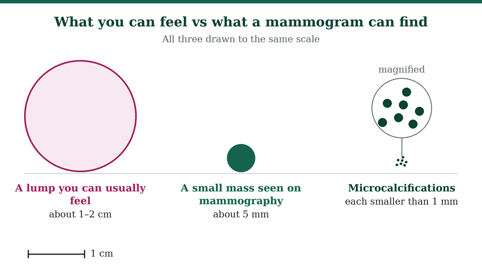
- Tiny clusters of microcalcifications, often smaller than a millimetre, which can be the first sign of ductal carcinoma in situ (DCIS), an early non-invasive cancer
- Small masses only a few millimetres across
- Subtle distortion in the pattern of the breast tissue
None of these can be felt. A lump that you or your doctor can feel is usually a centimetre or more. The only way to find the earlier changes is imaging good enough to show them.
Why small cancers hide: dense breasts
On a mammogram, fat looks dark, while dense glandular tissue and tumours both look white. In a dense breast, a small tumour can sit hidden behind normal tissue.
A landmark study in the New England Journal of Medicine found that women whose mammograms were 75% or more dense had nearly five times the odds of breast cancer being found at screening, or within a year of a negative screen, compared with women with very little density [2]. Much of that was due to cancers masked by dense tissue, and the effect was greater in younger women [2]. Dense breasts are common, which is one more reason the quality of the imaging matters.
What makes a mammogram good
Fortis Mohali's mammography system offers each of the capabilities below [8]. Each one solves a specific problem.
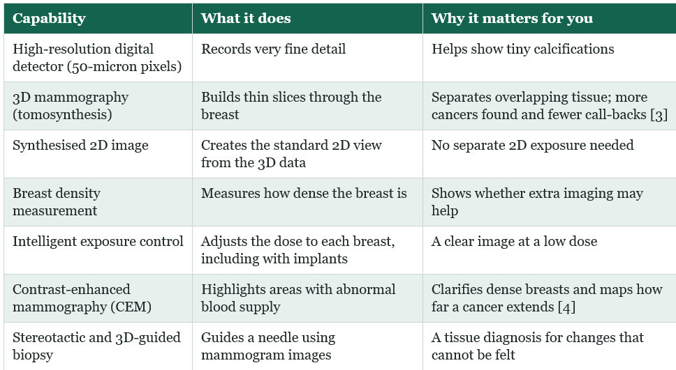
The evidence for 3D is strong. In more than 4.5 lakh screening mammograms across 13 US centres, adding 3D to standard digital mammography raised cancer detection from 4.2 to 5.4 per 1,000 women screened. Call-backs for extra tests fell from 107 to 91 per 1,000 [3].
For contrast-enhanced mammography, a 2022 meta-analysis of 19 studies found it detected breast lesions with sensitivity similar to breast MRI: 97% versus 96% [4]. That makes it a practical option for women who need contrast imaging but cannot have an MRI.
The machine is only half the story. Images need careful reading, and at Fortis Mohali every breast cancer case is reviewed by our tumour board before surgery is planned [6].
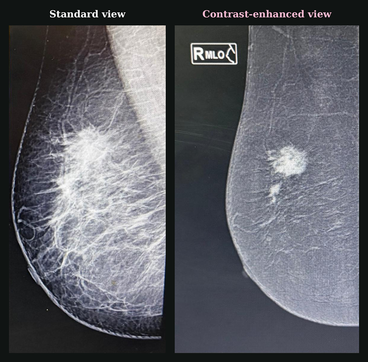
When a mammogram finds something you cannot feel: stereotactic biopsy
Finding a tiny change only helps if it can be tested. Microcalcifications and some small masses show up on a mammogram but cannot be felt, and are often invisible on ultrasound. For these, we use stereotactic biopsy.
The breast is gently compressed, as for a mammogram, and images taken from two angles pinpoint the exact spot. Under local anaesthetic, a needle is guided to it and small tissue samples are taken through a tiny nick in the skin. There is no surgical incision, and it is done as an outpatient procedure.
Image-guided needle biopsy accurately diagnoses most breast abnormalities, often faster and at lower cost than surgical biopsy, without leaving a cosmetic deformity [9]. If the result is benign, surgery is usually not needed. When a change is visible only on the 3D images, the biopsy can be guided using those slices instead.
Why routine screening deserves advanced technology too
Many people assume advanced imaging is only for women who already have a problem. I see it the other way round. Screening is where the smallest cancers are found. If the image cannot show a lesion a few millimetres across, a normal report is false reassurance, and the next chance to find it may be a year away.
Better imaging also means fewer unnecessary call-backs [3], which spares women anxiety and extra tests. A baseline 3D mammogram with a density report also tells you whether you need anything more.
Before your next mammogram, ask:
- Is the mammogram digital, and is 3D (tomosynthesis) available?
- Will my breast density be reported?
- Who will read my images?
- If something needs a closer look, are ultrasound, contrast-enhanced mammography and biopsy available in the same centre?
- Will my previous mammograms be compared? Bring them with you.
Breast self-examination: know your normal
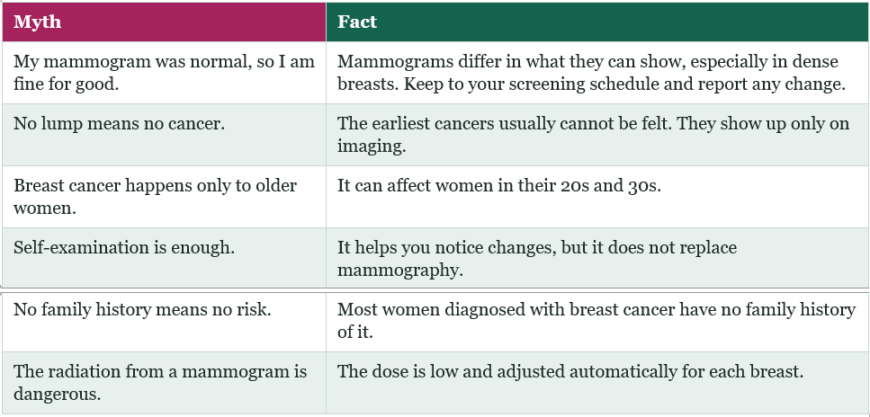
I want to be honest about self-examination. On its own, it is not a substitute for mammography. A Cochrane review of large trials found that routine self-examination, used as a screening method, did not reduce breast cancer deaths and led to more biopsies [5].
Knowing what is normal for your body still matters. The WHO puts awareness of warning signs and timely diagnosis at the centre of early detection [7]. Many women notice a change themselves, especially women under 40 and those between mammograms.
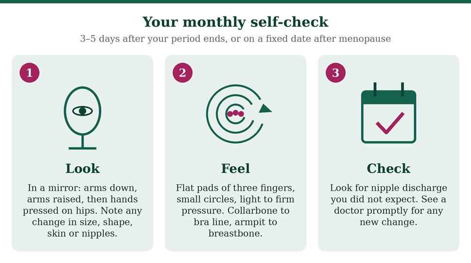
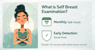
How to check, once a month:
- When: 3–5 days after your period ends. After menopause, pick a fixed date each month.
- Look: in front of a mirror, arms at your sides, then raised overhead, then hands pressed on your hips. Look for changes in size, shape, skin or nipples.
- Feel, lying down: right arm behind your head. With the flat pads of the three middle fingers of your left hand, move in small circles with light, medium and firm pressure. Cover the area from the collarbone to the bra line, and from the armpit to the breastbone. Repeat on the other side.
- Feel, in the shower: wet, soapy skin makes changes easier to notice.
- Check the nipples: note any discharge you did not expect.
See a doctor promptly if you notice a new lump in the breast or armpit, thickening or swelling, dimpling or puckering of the skin, a change in colour of the areola or flaky skin, a nipple that has turned inwards, or nipple pain or unexpected discharge. Most lumps turn out not to be cancer, but every new one deserves an examination.
When to start, and how often
- Women aged 40 and above: I advise a mammogram every year.
- Higher risk: a strong family history, a known BRCA mutation or previous chest radiation may mean starting earlier with a tailored plan. Speak to a breast specialist.
- Under 40, no symptoms: monthly breast awareness and a clinical breast examination at routine check-ups. For any symptom, see a breast specialist; ultrasound is usually the first test at this age.
Myth vs Fact
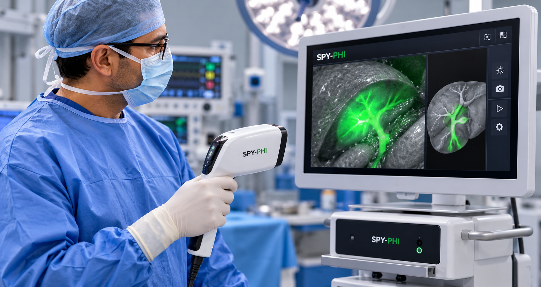
From detection to surgery: SPY-PHI fluorescence imaging
Precision does not stop at diagnosis. Our other new addition is in the operating theatre: the SPY-PHI fluorescence imaging system.
A small amount of a fluorescent dye, indocyanine green (ICG), is injected. A handheld near-infrared camera then shows it glowing on a screen in real time, alongside the normal view of the operating field [10]. In breast surgery, I use it for two things:
- Finding the sentinel lymph nodes. These are the first nodes that drain the breast, which we remove to check whether cancer has spread. A network meta-analysis of 35 studies found that ICG fluorescence identified sentinel nodes better than blue dye alone, with the lowest false-negative rate of the methods compared [11].
- Checking blood supply. During breast conservation with reconstruction, or mastectomy with reconstruction, it shows how well the skin and tissue are perfused. Its quantification software gives relative perfusion values and colour maps, an objective measure rather than the eye alone [10]. Reviews of the evidence show that fluorescence angiography typically reduces skin flap necrosis after mastectomy and reconstruction [12].
It is most valuable when blood supply is uncertain, and it supports the surgeon's judgement rather than replacing it.
What to expect at Fortis Mohali
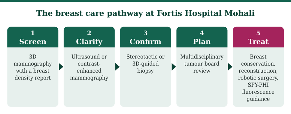
- Your mammogram, with 3D imaging where advised and a density report.
- If an area needs a closer look: ultrasound, contrast-enhanced mammography or an image-guided biopsy, usually within the same unit.
- If cancer is diagnosed: review by the multidisciplinary tumour board, then a plan that may include breast-conserving surgery, oncoplastic reconstruction, or robotic surgery for appropriately selected patients, with SPY-PHI fluorescence imaging used to map sentinel nodes and check blood supply.
Book a mammogram or a consultation with Dr. Naval Bansal: call 72728 72728
Categories
Clear allRelated Blogs
View all




FAQs
What is the difference between 2D and 3D mammography?
A 2D mammogram is one flat picture of the breast, so overlapping tissue can hide or mimic a lesion. 3D mammography takes images from several angles and builds thin slices, so the radiologist can look through the breast layer by layer.
Is 3D mammography safe?
Mammography uses a low dose of radiation, adjusted automatically for each breast. When a synthesised 2D image is used, a separate 2D exposure is not needed.
What is contrast-enhanced mammography, and who needs it?
An iodine-based contrast is injected into a vein, then two low-dose images are taken at different energies. Areas with abnormal blood supply, which cancers often have, stand out. It is used for dense breasts, unclear results and mapping a newly diagnosed cancer. You will need a kidney function test first, and it is not suitable in pregnancy or if you are allergic to iodine contrast.
I have been told I have dense breasts. What should I do?
Dense breasts are common and normal, but they can hide cancer and slightly raise risk. Ask your doctor whether 3D mammography, ultrasound or contrast-enhanced mammography should be added.
I am under 40. Do I need a mammogram?
Not routinely, unless you are at higher risk. Know what is normal for you and see a breast specialist for any change; ultrasound is usually the first test.
Does a mammogram hurt?
Compression is firm and lasts a few seconds per view. Most women find it uncomfortable rather than painful. Booking about a week after your period can help.
How should I prepare?
Avoid deodorant, talc or lotion on the day, wear a two-piece outfit, and bring any previous mammograms and reports.
What is a stereotactic biopsy, and does it hurt?
It is a needle biopsy guided by mammogram images, used for changes that cannot be felt or seen on ultrasound. It is done under local anaesthetic, so most women feel pressure rather than pain, and it needs only a tiny nick in the skin.
What is SPY-PHI fluorescence imaging?
It is a handheld near-infrared camera used during surgery. After a small injection of a fluorescent dye called indocyanine green, it shows lymph nodes and blood flow on a screen in real time. This helps the surgeon find the sentinel nodes and check that tissue has a healthy blood supply. Tell your team about any allergies, including to iodine.

Keep track of your appointments, get updates & more!

