
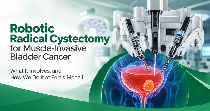


Robotic Radical Cystectomy for Muscle-Invasive Bladder Cancer - What It Involves and How We Do It at Fortis Mohali
By Dr Dharmender Aggarwal | Senior Consultant — Urology, Uro-Oncology & Robotic Surgery, Fortis Hospital, Mohali
MCh Urology (PGIMER, Chandigarh) | Fellowship in Robotic Surgery & Uro-Oncology, Royal College of Surgeons of England (St George’s University Hospital NHS Trust) | ERUS certified | ORSI certified | 950+ robotic cancer surgeries | Proctor and trainer for robotic urology programs

Very few conversations in my clinic are as difficult as the one that begins with the words “the cancer has gone into the muscle.” Patients hear “your bladder will have to come out” and stop hearing anything after that. If that is where you are right now, I want to slow the conversation down and explain what actually happens, what the surgery is trying to achieve, and what life looks like on the other side of it.
Understanding muscle-invasive bladder cancer
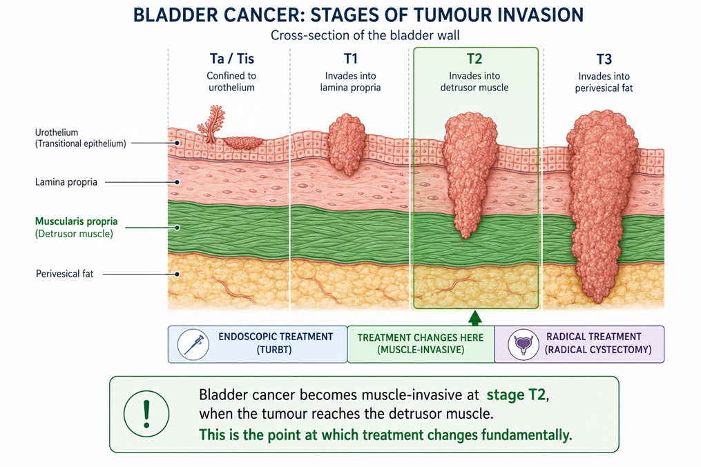
Bladder cancer starts in the inner lining of the bladder. While it stays in that lining, it can usually be managed endoscopically with TURBT and intravesical treatments such as BCG, with regular surveillance cystoscopies.
When the tumour grows into the detrusor muscle of the bladder wall, the disease behaves differently. It is classified as muscle-invasive bladder cancer, or MIBC, and it carries a real risk of spreading to the pelvic lymph nodes and beyond. Scraping the tumour away is no longer enough. This is the point at which definitive treatment becomes urgent rather than optional.
Bladder cancer is the ninth most commonly diagnosed cancer worldwide, with an estimated 614,298 new cases in 2022, and it is substantially more common in men than in women. In India, the National Cancer Registry Programme recorded 25,873 new cases in 2020 with a projected rise in the years since. In our own practice in Punjab, Himachal Pradesh and upper north India, tobacco use remains the dominant risk factor, followed by occupational exposure to aromatic amines and dyes.
The warning sign patients most often ignore
The most common presenting symptom is blood in the urine. It is frequently painless, it often appears once and then stops, and this is precisely why it gets dismissed. Other signs include increased urinary frequency, urgency, burning that does not settle with antibiotics, and pelvic discomfort.
Blood in the urine that resolves on its own is not reassurance that nothing is wrong. It is a reason which demands further investigations and a cystoscopy and imaging.
Why radical cystectomy remains the standard operation
For non-metastatic muscle-invasive bladder cancer, radical cystectomy — complete removal of the bladder together with an extended pelvic lymph node dissection, and the creation of a new pathway for urine — is the established standard of care in both European and American guidelines. For most patients, no other single modality matches it for long-term disease control.
I want to be precise about one thing here, because it is a distinction that gets blurred in a lot of health content:
Radical cystectomy with pelvic lymph node dissection is the standard cancer operation. Robotic assistance is the approach through which that operation is performed.
The robot does not change what is removed. It changes how the surgeon reaches it, how much trauma the body absorbs in the process, and how quickly the patient recovers afterwards. That distinction matters, and it is why the choice of centre matters more than the choice of machine.
The operation itself has two halves, and both are demanding:
- Complete cancer clearance — removal of the bladder with the prostate in men, or the uterus and anterior vaginal wall in women where indicated, together with an extended pelvic lymphadenectomy.
- Urinary reconstruction — building a new route for urine to leave the body using a segment of the patient’s own intestine.
Where robotic surgery fits, and what the evidence actually shows
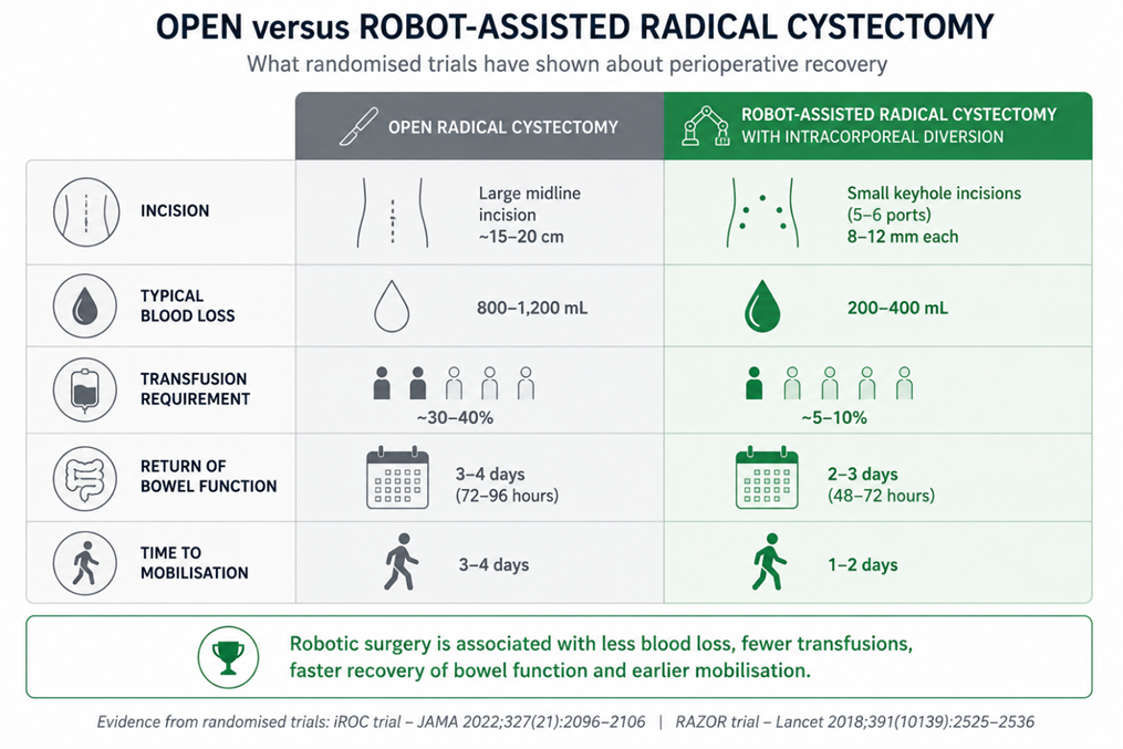
Open radical cystectomy is effective, but it is one of the most physically punishing operations in urology: a long midline incision, considerable blood loss, a prolonged period before the bowel starts working again, and weeks of recovery.
Robot-assisted radical cystectomy has been performed since 2003 and is now the preferred approach in most high-volume robotic centres. Two randomised trials give us the evidence base that patients should know about:
- The RAZOR trial (Lancet, 2018): robot-assisted radical cystectomy was non-inferior to open surgery for cancer recurrence at two years, with lower blood loss, fewer transfusions and shorter hospital stays. Reconstruction in that trial was done through an open incision.
- The iROC trial (JAMA, 2022): this compared robotic cystectomy with fully intracorporeal urinary diversion against open surgery across nine UK centres. Patients in the robotic arm spent significantly more days alive and out of hospital in the 90 days after surgery, with fewer complications and better early physical recovery.
Taken together, the evidence supports a straightforward statement: in experienced hands, the robotic approach delivers the same cancer operation with a lighter perioperative toll. It does not make the operation minor, and it does not compensate for inexperience.
What the technology gives the surgeon
- Magnified 3D vision: the neurovascular bundles, lymphatic channels and small pelvic vessels are visible with a clarity that is simply not available to the naked eye through an open incision.
- Wristed, tremor-filtered instruments: a full range of articulation deep in the narrow male pelvis, where the most demanding part of this dissection takes place.
- Reduced blood loss: most of the patients complete the surgery without needing a blood transfusion, which matters because perioperative transfusion has itself been linked to poorer outcomes in urological cancer surgery.
- Earlier return of bowel function: the bowel is handled less and exposed less, so patients typically start oral fluids and mobilise sooner.
- A thorough lymph node template: precision at depth supports a properly extended nodal dissection, which is an important part of the oncological quality of this operation.
- Lesser catabolism and strain on body: Robotic technique allows surgeon to perform surgery with lesser strain on body’s physiological processes and less catabolism. This allows surgery to be performed in patients with many comorbidities and with lesser overall complications.
Urinary diversion: the decision that shapes your daily life
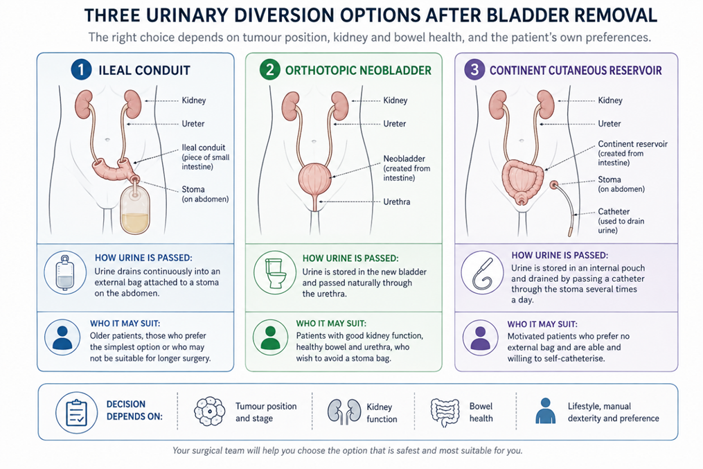
Once the bladder is removed, urine needs a new route out. This is the part of the conversation that patients think about most, and rightly so, because it affects everyday living far more than the cancer surgery itself does.
Ileal conduit
A short segment of small intestine carries urine to a small opening on the abdominal wall, where it collects in a discreet external appliance. It is the most widely performed diversion, it is reliable, and it involves the least metabolic demand on the body. Stoma training begins before discharge and most patients are managing independently within a few days.
Orthotopic neobladder
A new reservoir is fashioned from a segment of intestine and joined directly to the natural urinary passage, allowing urine to be passed in the usual way without an external appliance. Continence improves progressively over one to three months with pelvic floor training. It suits motivated, well-selected patients with suitable tumour position, adequate kidney function and healthy bowel.
Continent cutaneous reservoir
An internal pouch is created and emptied at intervals through a small, continent abdominal opening using a catheter. It is an option for selected patients for whom a neobladder is not suitable but who wish to avoid an external appliance.
There is no diversion that is best for everybody. Age, overall health, comorbidities, kidney function, bowel health, tumour location, dexterity, independence and personal preference all feed into it. We always spend an extra consultation on this decision than have a patient discover six months later that they were not told what to expect.
Why we do the reconstruction inside the body
At Fortis Mohali, Dr Aggarwal’s team perform intracorporeal urinary diversion, meaning the conduit or neobladder is constructed entirely within the abdomen using the robotic instruments, rather than by delivering bowel out through a larger incision. This avoids that additional incision, reduces fluid and heat loss from exposed bowel, and is associated with the smoother recovery. It allows faster recovery of bowel function and greatly decreases hospital stay. It is technically the most demanding version of this surgery and it is appropriate only where a program does enough of these cases to do them well. It is always recommended to have these complex surgeries performed at a centre which does it on routine basis.
What the pathway looks like at Fortis Mohali
This surgery is protocol-driven from the first consultation. Nothing about it should feel improvised.
- Staging and review: CT urography or MRI, a review of the original TURBT biopsies, and assessment of kidney, cardiac, pulmonary and nutritional status.
- Multidisciplinary tumour board: Uro-oncology, medical oncology, radiation oncology, radiology and pathology discuss the case together. Not every patient should go straight to surgery — some need cisplatin-based chemotherapy first, and a few are better served by a bladder-preserving trimodal approach.
- Pre-operative counselling and stoma siting: diversion options are discussed with you and your family, and where a stoma is planned, the site is marked in advance by a trained stoma therapist with you sitting, standing and bending.
- Surgery: performed on the da Vinci Xi platform through five to six 8 mm ports, with intracorporeal reconstruction.
- Enhanced Recovery After Surgery (ERAS): structured pain control, early mobilisation, graded reintroduction of diet, thromboprophylaxis and chest physiotherapy, with round-the-clock critical care cover available.
- Structured follow-up: final pathology review, a decision on any further treatment, monitoring of kidney function and electrolytes, surveillance imaging, and ongoing diversion, nutritional and rehabilitation support.
Life after bladder removal
Patients ask me whether they will live normally afterwards. For most, the answer is yes, with an adjustment period. Stoma care is usually learned within a few days. Neobladder continence builds over one to three months with pelvic floor work. Many patients are back to desk-based work within a few weeks and to fuller activity over roughly six weeks, though this varies considerably with age, nutrition and overall recovery.
Follow-up is not optional. Kidney function, electrolytes, blood gas analysis and surveillance imaging all need monitoring long after the surgery is behind you.
Myth vs Fact
These are the six misconceptions I correct most often in clinic.
Robotic surgery means the robot operates on me.
The robotic system has no autonomy. Every movement of every instrument is directed in real time by the surgeon at the console. The platform provides magnified 3D vision and wristed instruments; the judgement and the dissection remain entirely human. Robotic machine only works as an advanced instruments in the hand of experienced surgeons.
Keyhole surgery means bladder cancer is only partly removed.
The operation performed is the same cancer operation: complete removal of the bladder along with an extended pelvic lymph node dissection. Randomised evidence has found cancer outcomes same to open surgery when the procedure is done by experienced teams. All steps include maintaining intact bladder removal without opening or spilling any cancer cells.
Once the bladder is removed, I will have to live with a bag forever.
Not necessarily. An ileal conduit with a stoma is one option. Selected, well-counselled patients can be offered an orthotopic neobladder, which allows urine to pass through the natural passage. Suitability depends on tumour location, kidney function, bowel health and how much self-care a patient can commit to.
Robotic surgery is a small procedure with a quick fix.
Radical cystectomy is major surgery regardless of the approach. It takes around 4 hours of surgery. Smaller incisions reduce blood loss and speed early recovery, but the operation still needs careful pre-assessment, ICU-level monitoring where required, and structured follow-up.
Blood in urine that stops on its own is nothing to worry about.
Painless, intermittent blood in the urine is the single most common presenting sign of bladder cancer. It stopping is not reassurance. It warrants an evaluation.
Why consider Fortis Hospital, Mohali
Robotic cystectomy with intracorporeal diversion is not a procedure to be attempted occasionally. It needs volume, a full multidisciplinary structure, and a team that has managed all aspects of recovery including the complications as well as the successes.
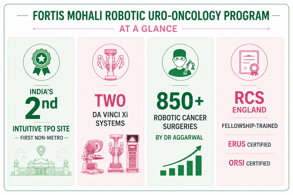
- An Intuitive Total Program Observation site: Fortis Mohali has been designated by Intuitive Surgical as India’s second Total Program Observation (TPO) site, and the first outside a metro city. TPO sites host visiting surgical teams from other institutions who come to observe an established robotic program in operation. It is an external assessment of the program, not a claim we make about ourselves.
- Two da Vinci Xi systems: the fourth-generation da Vinci Xi platform, with a second console added to support the volume of the program across specialties.
- A dedicated uro-oncology setup: robotic theatre with frozen section availability, uro-pathology, medical oncology for neoadjuvant and adjuvant therapy, radiation oncology, and interventional radiology within the same campus.
- Trained stoma and uro-oncology nursing: dedicated stoma therapists who counsel the patient and the family, not just the patient, along with physiotherapy and clinical nutrition support through the recovery period.
- Surgeon experience: Dr Dharmender Aggarwal have performed over 950 robotic cancer surgeries of the kidney, bladder, prostate and upper urinary tract, trained through the Royal College of Surgeons of England, and he proctor and train surgeons setting up robotic programs at other institutions. He is a senior consultant with dedicated approach to Urology cancer cases and a high-volume robotic surgeon.
In this operation, millimetres decide margins, continence and potency. The platform provides the control. The outcome still depends on the person at the console and the team around the patient for the fortnight that follows.
When to seek a specialist opinion
Please come and see us if you have:
- Been diagnosed with muscle-invasive or high-grade bladder cancer
- Been advised open bladder removal and want to know whether a robotic approach with intracorporeal diversion is possible in your case
- High-grade non-muscle-invasive disease that has not responded to BCG
- Blood in the urine with muscle invasion reported on your TURBT
- Been told the bladder must come out, and want to understand your diversion options before you decide
Please bring your cystoscopy findings, TURBT histopathology slides and blocks, CT or MRI films, kidney function reports and details of any treatment already received. Reviewing the actual images and slides tells me far more than a summary letter does. We see patients referred from across Punjab, Haryana, Himachal Pradesh and Jammu & Kashmir.
Book a uro-oncology consultation
Dr Dharmender Aggarwal — Urology, Uro-Oncology & Robotic Surgery
Fortis Hospital, Sector 62, Phase VIII, Mohali, Punjab
Call 72728 72728
A bladder cancer diagnosis rewards decisive assessment. If this operation is done correctly the first time, it offers the best chance of cure with the least lasting toll on your body.
Categories
Clear allMeet the doctor

- Urology | Urology | Uro-Oncology
-
 13 Years
-
 1050
1050
Related Blogs
View all



Adult Circumcision: Debunking Facts and Myths | Fortis Health Connect Blogs
Is Beer Good for Kidney Stones ? - Myths Vs Facts | Fortis Healthcare




FAQs
Is robotic radical cystectomy the standard treatment for muscle-invasive bladder cancer?
Radical cystectomy with pelvic lymph node dissection is the established standard operation for non-metastatic muscle-invasive bladder cancer. Robotic assistance is the surgical approach through which that standard operation is delivered. In experienced, high-volume centres it has become the preferred approach because randomised trials have shown lower blood loss, fewer transfusions and faster early recovery, with comparable cancer control.
How long will I stay in hospital?
This varies with the diversion performed, your fitness and how your bowel recovers. Many patients undergoing robotic cystectomy with an enhanced recovery protocol are mobilised within a day and discharged appreciably sooner than after open surgery. Your team will give you a realistic estimate for your specific case at the pre-operative counselling session.
What is intracorporeal urinary diversion and why does it matter?
It means the new urinary pathway, whether an ileal conduit or a neobladder, is constructed entirely inside the abdomen using the robotic instruments, rather than by pulling the bowel out through an incision. It avoids a larger opening, reduces fluid and heat loss from exposed bowel, and is associated with a smoother early recovery. It is technically demanding and is offered where the team has the volume and experience for it.
Can I pass urine normally after my bladder is removed?
Selected patients can be offered an orthotopic neobladder, constructed from a segment of intestine and joined to the natural urinary passage, allowing urine to be passed in the usual way. Continence typically improves over one to three months with pelvic floor training. Not everyone is a candidate; tumour position, kidney function, bowel condition and a patient’s ability to manage self-
Will I need chemotherapy as well as surgery?
Some patients with muscle-invasive disease benefit from cisplatin-based chemotherapy before surgery, and some need further treatment afterwards depending on the final pathology. This is decided jointly by the uro-oncology team, surgeon and the medical oncologist after assessing kidney function, fitness and other conditions.
I have been advised open surgery elsewhere. Is a second opinion reasonable?
Yes, and it is common. Bring your cystoscopy findings, TURBT histopathology records, CT or MRI imaging, kidney function reports and details of any treatment already received. A review of the actual images and slides is far more useful than a summary letter.
How soon can I get back to work?
Most patients return to desk-based work within a few weeks and to fuller activity over roughly six weeks, though this depends on the diversion, age, nutrition and any complications. Stoma or neobladder training begins before discharge so that you are not learning it alone at home.

Keep track of your appointments, get updates & more!

